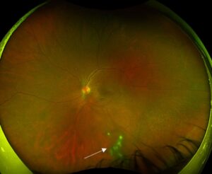
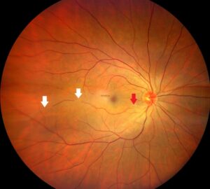
A macular hole retinal detachment is a detachment of the posterior pole caused by a full-thickness hole at the fovea.
It occurs mainly in highly myopic eyes with a posterior staphyloma.
Therefore, it behaves differently from a typical rhegmatogenous detachment and needs a tailored plan.
How Macular Hole Retinal Detachment Differs
Most detachments start from peripheral tears.
This type starts at the macula and spreads outward.
Also, the detachment may stay confined to the posterior pole for months.
As a result, the patient notices central loss and not a curtain.
Why High Myopia Predisposes
Long eyes stretch the retina and thin the choroid.
A posterior staphyloma creates a concave posterior wall that adds traction.
Also, retinoschisis at the macula often precedes the hole.
Therefore, the retina separates easily from the pigment epithelium.
Fundus Explorer Pro
Photograph the retinal findings described here with the phone already in your pocket — 22 D optics and built-in illumination in one handheld unit.
From Choroida — the team behind this siteMechanism of Macular Hole Retinal Detachment
Morita and colleagues showed that axial length and staphyloma were key causes.
Fluid enters through the hole and tracks under the retina.
Also, the stretched, stiff retina resists reattachment.
As a result, the pump of the pigment epithelium may fail to dry the space.
Risk Factors for Macular Hole Retinal Detachment
Axial length above 30 millimeters and a large staphyloma raise the risk.
Chorioretinal atrophy and foveal schisis add to it.
Also, older age and prior vitrectomy play a role.
In addition, trauma can precipitate the hole.
Symptoms of Macular Hole Retinal Detachment
Patients report a central scotoma and distortion.
Vision falls to counting fingers in advanced cases.
Also, peripheral vision stays normal until late.
Therefore, symptoms may be blamed on myopic maculopathy.
Diagnosing Macular Hole Retinal Detachment on OCT
OCT shows a full-thickness hole with subretinal fluid in a posterior detachment (see retinal detachment in high myopia).
The scleral curve can be extreme.
Also, schisis layers may be visible.
Therefore, OCT confirms the diagnosis and helps plan surgery.
Ultrasound and Wide-Field Imaging
B-scan shows the staphyloma and the detached retina in poor media.
Wide-field OCT captures the posterior curve in one scan.
Also, it helps to find peripheral tears.
As a result, imaging should look for both macular and peripheral breaks.
Surgical Goals
The aims are to relieve traction, close the hole and flatten the retina.
Surgery must also address the staphyloma in some eyes.
Also, the choice depends on the extent of the detachment.
Therefore, plan the approach before the operation.
Vitrectomy with Membrane Peeling
Standard vitrectomy removes the vitreous cortex and the internal limiting membrane.
Gas or silicone oil provides tamponade.
Also, the results are poorer than in non-myopic holes.
As a result, many surgeons add other steps.
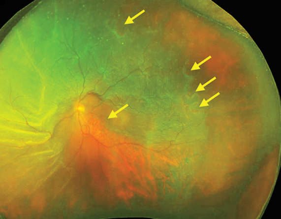
ILM Flap Technique
Kuriyama and colleagues reported better closure with an inverted flap in myopic holes.
The flap helps seal the hole and bridges the gap (see ILM flap technique).
Also, success rates exceed those of simple peeling in many series.
Therefore, the flap is now a common choice.
Macular Buckling
A macular buckle presses the sclera forward to meet the retina.
It addresses the staphyloma directly.
Also, Ripandelli and colleagues compared vitrectomy and posterior buckling and found good results with the buckle.
In addition, it is technically demanding.
Choosing the Tamponade
Silicone oil gives long support and is useful in large detachments.
Gas allows quicker visual recovery.
Also, face-down positioning is hard in myopic patients with kyphosis.
Therefore, individual factors guide the choice.
Outcomes of Macular Hole Retinal Detachment Surgery
Anatomic success ranges widely between series.
Visual outcome depends on atrophy and the duration of detachment.
Also, many eyes keep poor central vision despite closure.
As a result, set realistic goals.
Complications
Hole reopening and recurrent detachment occur.
Epiretinal membrane and cataract follow vitrectomy.
Also, macular atrophy may progress.
In addition, silicone oil carries its own risks.
Counseling Patients
Explain the lower success rates compared with other holes.
Describe the possibility of more than one operation.
Also, discuss the limits of vision recovery.
Therefore, shared decisions are important.
Pitfalls to Avoid
- Do not treat this detachment as a simple macular hole
- Never forget to look for peripheral tears
- Do not use gas alone in a large detachment without a plan
- Never ignore the staphyloma when choosing surgery
Role of Preoperative Imaging
Wide-field OCT shows the shape of the posterior wall.
It helps decide between a flap and a buckle.
Also, it measures hole size and the depth of detachment.
Therefore, imaging guides the surgical plan.
Postoperative Follow-Up
Check OCT at one week, one month and three months.
Look for hole closure, residual fluid and the flap.
Also, monitor for epiretinal membrane.
In addition, refer patients early for low-vision support.
For the latest studies, see the PubMed results for macular hole retinal detachment.


Document what you see
Two smartphone imaging tools built for everyday clinic use — one for the slit lamp, one for the fundus.
From Choroida — the team behind this siteReferences
- Morita H, Ideta H, Ito K, Yonemoto J, Sasaki K, Tanaka S. Causative factors of retinal detachment in macular holes. Retina. 1991;11:281-284.
- Ripandelli G, Coppé AM, Fedeli R, Parisi V, D’Amico DJ, Stirpe M. Evaluation of primary surgical procedures for retinal detachment with macular hole in highly myopic eyes: a comparison of vitrectomy versus posterior episcleral buckling surgery. Ophthalmology. 2001;108:2258-2264.
- Kuriyama S, Hayashi H, Jingami Y, Kuramoto N, Akita J, Matsumoto M. Efficacy of inverted internal limiting membrane flap technique for the treatment of macular hole in high myopia. Am J Ophthalmol. 2013;156:125-131.
- Ohno-Matsui K. Pathologic myopia. Asia Pac J Ophthalmol. 2016;5:415-423.

Writes and reviews the clinical articles on Educate. Every post is checked against peer-reviewed sources before it goes live.