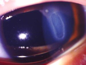
Stocker’s line is a fine, brown-yellow line of iron in the corneal epithelium that runs in front of the head of a pterygium.
It marks the advancing edge of the lesion on the cornea, so a careful look at the line helps you judge how long the pterygium has been there and how active it is.
What Stocker’s Line Represents
Iron accumulates in basal epithelial cells where the surface is uneven and the tear film pools. The line follows the curve of the pterygium head and sits just beyond its apex, and because the deposit lies in the epithelium, it does not cause symptoms of its own.
How to Find Stocker’s Line
Use the slit lamp with the cobalt blue filter, because the iron stands out as a dark line. Look in the clear cornea just beyond the apex of the head, and the line may be partial or faint, so scan slowly. Teachers often take the line as a sign of a slowly growing lesion, although the evidence is limited.
All-fit Slit-Lamp Adapter
Record and share exactly what you see at the slit lamp. One adapter fits any slit lamp or surgical microscope — and any smartphone.
From Choroida — the team behind this siteStocker’s Line Versus Other Iron Lines
Location tells you the cause, for example the Hudson-Stahli line, which is horizontal and sits in the lower cornea of older people. The Fleischer ring surrounds the cone in keratoconus, and Ferry’s line sits in front of a filtering bleb. Therefore, a line at the head of a pterygium is Stocker’s line (see corneal iron lines).
Managing the Pterygium
The line itself needs no treatment, so manage the pterygium with lubricants and ultraviolet protection when it is small and quiet (see pterygium). Consider excision with a conjunctival autograft if the lesion threatens the visual axis, causes astigmatism or stays inflamed (see conjunctival autograft).
History and Symptoms in Pterygium
Ask about foreign body sensation, redness, dryness and blur from induced astigmatism, and also ask about sun exposure, wind, dust and outdoor work. Because the lesion grows slowly, many patients report years of symptoms.
Features of the Pterygium Head
Look at the head for small gray islands called Fuchs islets, and check the cornea for induced astigmatism with topography. A thick, fleshy lesion with prominent vessels is more active than a flat, pale one.
Pseudopterygium
In contrast, a pseudopterygium is a scar band attached to the cornea after injury or inflammation, and a probe can pass under its neck, which differs from a true pterygium. Therefore, ask about previous burns, ulcers or surgery.
Surgical Considerations
Recurrence is the main problem after excision, and conjunctival autograft gives lower recurrence rates than bare sclera excision. Patients should also avoid sun and wind exposure after surgery to lower the risk.
A Pitfall to Avoid
Ocular surface squamous neoplasia can mimic a pterygium, so send excised tissue for histology when the lesion looks atypical (see ocular surface squamous neoplasia) and watch for rapid growth, leukoplakia and prominent feeder vessels.
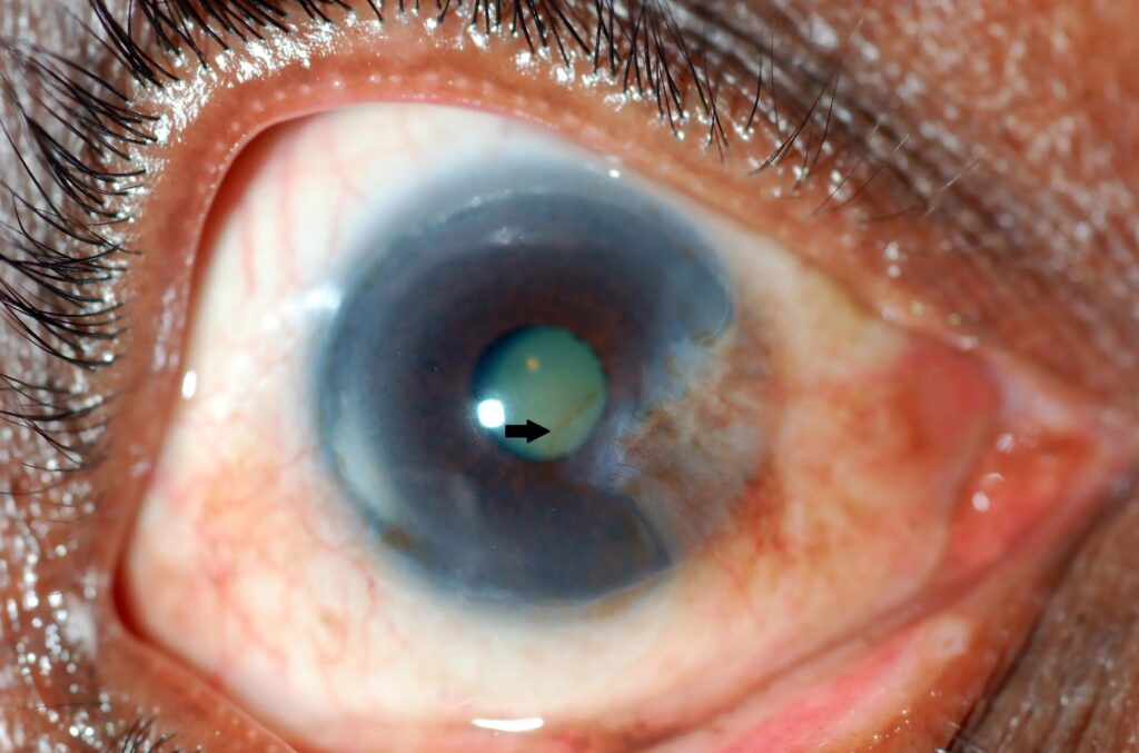
Reading the Line Together With the Lesion
A well-defined line suggests a stable head, and a blurred or absent line fits an active, advancing lesion. This reading is a clinical impression, so confirm it by following the lesion over time and photograph the head to compare at later visits.
Staining Pattern
A pterygium head may take fluorescein at its margin because the surface is uneven, while iron lines themselves do not stain because they lie within the epithelium. Therefore, use blue light without dye to see the line.
Symptoms That Need Attention
Pain, marked redness or a sudden change in the lesion call for review, and reduced vision from astigmatism or from encroachment on the visual axis also calls for referral.
Patient Counselling
Explain that the lesion is benign but can grow, and ask the patient to wear sunglasses and a hat outdoors and use lubricants for comfort. Because recurrence follows surgery in some patients, discuss it before the operation.
Role of Topography
Pterygia flatten the cornea along the axis of the lesion and induce astigmatism, and the effect grows as the head moves toward the visual axis. Therefore, measure topography before surgery and again after healing.
Stocker’s Line in Established Lesions
The line usually appears in long-standing lesions in adults who have spent years outdoors, while young patients with a small pterygium rarely show it. Because iron needs time to accumulate, the line points to a lesion that has been present for a while.
Photographing Stocker’s Line
Cobalt blue illumination gives the best photographs, and a smartphone adapter on the slit lamp captures them easily. Also, take a second image with white light so you can compare the pterygium head with the line.
Stocker’s Line and Surgical Planning
The line lies just beyond the head, so it gives a rough landmark for the corneal extent of the lesion, and you should record the distance from the limbus to the apex before surgery. Because the apex moves toward the visual axis over time, this number guides the timing of excision.


Document what you see
Two smartphone imaging tools built for everyday clinic use — one for the slit lamp, one for the fundus.
From Choroida — the team behind this siteReferences
- Gass JDM. The iron lines of the superficial cornea: Hudson-Stahli line, Stocker’s line, and Fleischer’s ring. Arch Ophthalmol. 1964;71:348-358.
- Chui J, Di Girolamo N, Wakefield D, Coroneo MT. The pathogenesis of pterygium: current concepts and their therapeutic implications. Ocul Surf. 2008;6:24-43.
- Hirst LW, Axelsen RA, Schwab I. Pterygium and associated ocular surface squamous neoplasia. Arch Ophthalmol. 2009;127:31-32.
- Mannis MJ, Holland EJ, eds. Cornea. 5th ed. Elsevier; 2022.
Test yourself
A few questions straight from this article.
-
What is Stocker's line?
Stocker's line is a fine brown-yellow line of iron in the corneal epithelium running in front of a pterygium head, marking its advancing edge. -
Why does iron accumulate to form Stocker's line ahead of a pterygium?
Iron accumulates in basal epithelial cells where the surface is uneven and the tear film pools, following the curve of the head just beyond its apex. -
Which slit lamp technique best shows Stocker's line?
With the cobalt blue filter the iron stands out as a dark line in the clear cornea just beyond the apex; the line may be partial or faint, so scan slowly. -
Which corneal iron line surrounds the cone in keratoconus?
Location tells the cause: the Fleischer ring surrounds the cone in keratoconus, the Hudson-Stahli line is horizontal in the lower cornea, and Ferry's line sits before a filtering bleb. -
Why does Stocker's line not stain with fluorescein?
The pterygium head may stain at its uneven margin, but iron lines do not stain because they lie within the epithelium, so blue light without dye is used. -
When should excision with a conjunctival autograft be considered for a pterygium with Stocker's line?
The line itself needs no treatment; a small quiet pterygium is managed with lubricants, and excision with autograft is considered if it threatens the visual axis, causes astigmatism or stays inflamed. -
Which pterygium operation gives lower recurrence rates than bare sclera excision?
Recurrence is the main problem after excision, and conjunctival autograft gives lower recurrence rates than bare sclera excision. -
Which examination finding distinguishes a pseudopterygium from a true pterygium?
A pseudopterygium is a scar band attached to the cornea after injury or inflammation, and a probe can pass under its neck, unlike a true pterygium. -
Which condition can mimic a pterygium and justifies histology of atypical excised tissue?
Ocular surface squamous neoplasia can mimic a pterygium, so atypical tissue goes for histology; watch for rapid growth, leukoplakia and prominent feeder vessels. -
In which patients is Stocker's line usually found?
Because iron needs time to accumulate, the line usually appears in long-standing lesions in adults who have spent years outdoors, and young patients rarely show it.

Writes and reviews the clinical articles on Educate. Every post is checked against peer-reviewed sources before it goes live.