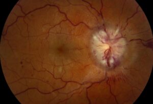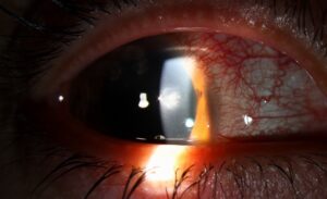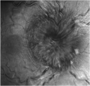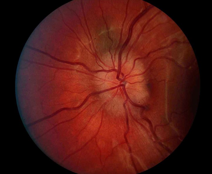
Optic disc swelling, also called optic disc edema, is an elevation and blurring of the optic nerve head.
It can come from raised intracranial pressure, local inflammation, ischemia, compression, or a vein occlusion.
Also, papilledema is the narrow term for swelling caused by raised intracranial pressure, always bilateral in its established form, and every other cause belongs under the broader heading.
The first job at the slit lamp is to decide whether the disc is truly swollen.
The second is to decide how urgent the cause is.
Optic Disc Swelling Versus Papilledema
Calling every swollen disc papilledema leads to unnecessary lumbar punctures in some patients and missed diagnoses in others (see is every optic disc swelling papilledema).
Papilledema develops over days to weeks, usually leaves central acuity intact until late, and enlarges the blind spot.
Also, swelling from an anterior optic neuropathy or optic neuritis is usually unilateral and affects acuity, color vision, or the field early.
Examination Findings in Optic Disc Swelling
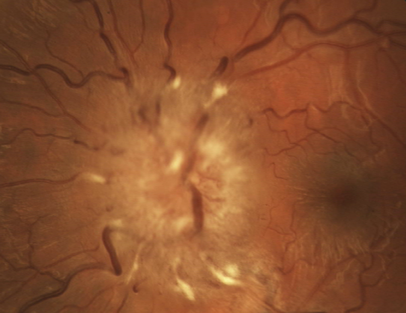
True swelling shows a blurred disc margin, elevation of the surface, hyperemia, and obscured vessels where they cross the edge of the disc.
Peripapillary hemorrhages, cotton-wool spots, venous engorgement, and absent spontaneous venous pulsations all support swelling, although about one in five normal eyes lack venous pulsations.
Choroidal folds, Paton lines, and a circumferential halo help grade severity.
The Frisén scale from 0 to 5 gives a repeatable record (see the references).
Fundus Explorer Pro
Photograph the retinal findings described here with the phone already in your pocket — 22 D optics and built-in illumination in one handheld unit.
From Choroida — the team behind this siteOptic Disc Swelling: Causes by Mechanism
- Raised intracranial pressure: mass lesions, venous sinus thrombosis, and idiopathic intracranial hypertension (see idiopathic intracranial hypertension)
- Ischemic: non-arteritic and arteritic anterior ischemic optic neuropathy (see non-arteritic anterior ischemic optic neuropathy)
- Inflammatory: papillitis from optic neuritis, sarcoid, and uveitis (see optic neuritis)
- Vascular: central retinal vein occlusion, papillophlebitis, and diabetic papillopathy
- Systemic: malignant hypertension, severe anemia, and hypotony
- Compressive and infiltrative: orbital tumors, glioma, lymphoma, and leukemia
Pseudo-Edema
Some discs look swollen and are not.
Optic disc drusen, a tilted disc, a crowded hyperopic nerve, and myelinated fibers can also all mimic edema.
The vessels are not obscured in the same way (see optic disc drusen).
In addition, autofluorescence and B-scan ultrasound show buried drusen, and fluorescein angiography shows leakage with true edema and none with pseudo-edema.
OCT of the retinal nerve fiber layer and the optic nerve head also helps to separate the two.
A hyporeflective shell around the disc favors edema.
Clues to the Cause
A bilateral swollen disc with headache, transient visual obscurations, and a normal acuity points toward raised pressure.
In contrast, a sudden, painless, sectoral field loss in a patient with a crowded disc and vascular risk factors suggests NAION, whereas pain on eye movement in a young adult suggests optic neuritis.
Jaw claudication, scalp tenderness, and a raised ESR in a patient over 50 call for emergency steroids for giant cell arteritis.
Flame hemorrhages in all quadrants with dilated veins suggest a vein occlusion (see central retinal vein occlusion).
Workup for Optic Disc Swelling
Measure the blood pressure, test the acuity, color vision, pupils, and fields, and check for a relative afferent pupillary defect.
Bilateral swelling with no clear cause needs urgent neuroimaging, preferably MRI with venography, followed by lumbar puncture with an opening pressure when imaging is normal.
Unilateral swelling needs an ESR, CRP, and platelet count in older patients, and MRI of the orbits and brain with contrast when the course is atypical.
Management and Follow-Up
Treatment follows the cause, and the swelling itself is not treated.
Also, raised intracranial pressure may need weight loss, acetazolamide, shunting, or optic nerve sheath fenestration to protect vision (see optic nerve sheath fenestration).
Follow the disc with OCT and photographs, and check the field regularly, since chronic swelling can end in optic atrophy with a pale disc and permanent loss.
An Example Case
A 29-year-old woman with obesity and daily headaches reports that her vision “grays out” when she bends over.
Acuity is 20/20 in both eyes, the pupils are equal, and both discs are swollen with a Frisén grade 3 and an enlarged blind spot on the field.
MRI and MR venography are normal, the opening pressure is 38 cm of water.
The diagnosis is idiopathic intracranial hypertension, treated with weight loss and acetazolamide.
Pitfalls to Avoid
- Do not label every swollen disc as papilledema before checking for a relative afferent pupillary defect
- Never delay steroids in a patient with suspected giant cell arteritis while waiting for a biopsy
- Do not forget the blood pressure, since malignant hypertension is an easily missed cause
- Never dismiss a pale disc with a history of swelling, as this may be optic atrophy from prolonged edema
For the latest studies, see the PubMed results for optic disc edema.


Document what you see
Two smartphone imaging tools built for everyday clinic use — one for the slit lamp, one for the fundus.
From Choroida — the team behind this siteReferences
- Hayreh SS. Pathophysiology of optic disc edema in raised intracranial pressure. Prog Retin Eye Res. 2016;50:108-144.
- Frisén L. Swelling of the optic nerve head: a staging scheme. J Neurol Neurosurg Psychiatry. 1982;45:13-18.
- Friedman DI, Liu GT, Digre KB. Revised diagnostic criteria for the pseudotumor cerebri syndrome in adults and children. Neurology. 2013;81:1159-1165.
- American Academy of Ophthalmology. Basic and Clinical Science Course, Section 5: Neuro-Ophthalmology. AAO; current edition.

Writes and reviews the clinical articles on Educate. Every post is checked against peer-reviewed sources before it goes live.