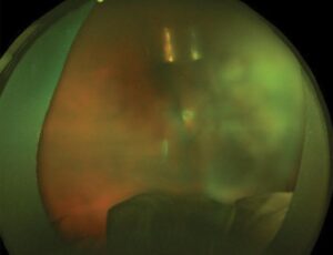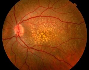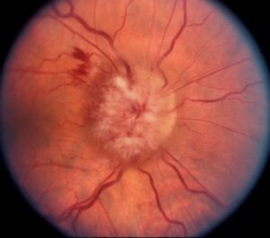
Modern retinal detachment repair has more good options than at any time in the past.
All methods aim to find and close every break, and to keep the retina attached while the seal forms.
Also, the choice depends on the type of detachment, the lens, and the surgeon’s skill, and new trial data have refined it.
The Goals of Modern Retinal Detachment Repair
- Identify all retinal breaks, which are the cause of the detachment
- Relieve traction at the breaks, with a buckle or by removing the vitreous
- Seal the breaks with laser or cryotherapy (see laser retinopexy and retinal cryotherapy)
- Support the retina with a gas bubble, buckle, or oil until the scar forms
Modern Retinal Detachment Repair: Pneumatic Retinopexy
A gas bubble is injected into the vitreous in the office, and the patient is positioned so that the bubble closes the break, followed by laser or cryotherapy.
It suits a single break or a cluster of breaks in the upper retina, with a clear view (see pneumatic retinopexy).
The PIVOT trial compared it with vitrectomy, and found that single-surgery success was lower with pneumatic retinopexy, but visual outcomes were better, with less distortion, in eyes treated that way (see the references).
Fundus Explorer Pro
Photograph the retinal findings described here with the phone already in your pocket — 22 D optics and built-in illumination in one handheld unit.
From Choroida — the team behind this siteScleral Buckle
A silicone band is sewn to the outside of the sclera, indenting the wall against the break.
- Good for young phakic patients with attached vitreous, lower breaks, or dialysis (see scleral buckle surgery)
- Avoids cataract formation, which is common after vitrectomy
- Requires skill, and can cause double vision, myopia, and discomfort
Modern Retinal Detachment Repair: Vitrectomy
Small-gauge vitrectomy removes the vitreous, releases traction, and uses gas or oil to flatten the retina.
- Best for pseudophakic eyes, large or multiple breaks, vitreous hemorrhage, and complex or proliferative disease (see small-gauge vitrectomy)
- Causes cataract in phakic eyes
- Needs positioning after surgery
The SPR study showed that vitrectomy tended to do better than scleral buckling in pseudophakic eyes, while buckling did well in phakic eyes (see the references).
Combined Methods
Some eyes need a buckle and vitrectomy together, especially with proliferative vitreoretinopathy or giant tears (see giant retinal tear).
Modern Retinal Detachment Repair: New Tools That Help
- Wide-angle viewing and heads-up display improve visualization of the periphery (see heads-up 3D vitreoretinal surgery)
- Heavy liquids flatten the retina, and long-acting gases and silicone oil provide support
- Intraoperative OCT checks the macula (see intraoperative OCT in retinal surgery)
- Research continues on prevention of proliferative vitreoretinopathy
Choosing for Each Patient
- Young phakic patient with a single upper break: pneumatic retinopexy or buckle
- Phakic patient with inferior or multiple breaks: buckle, sometimes combined
- Pseudophakic patient: vitrectomy in most cases
- Detachment with proliferative changes or hemorrhage: vitrectomy, with or without buckle
Counselling
Explain the options, and that the success rate with one operation is high, but that some patients need a second.
Tell patients to report new flashes, floaters, or a shadow without delay.
An Example Choice
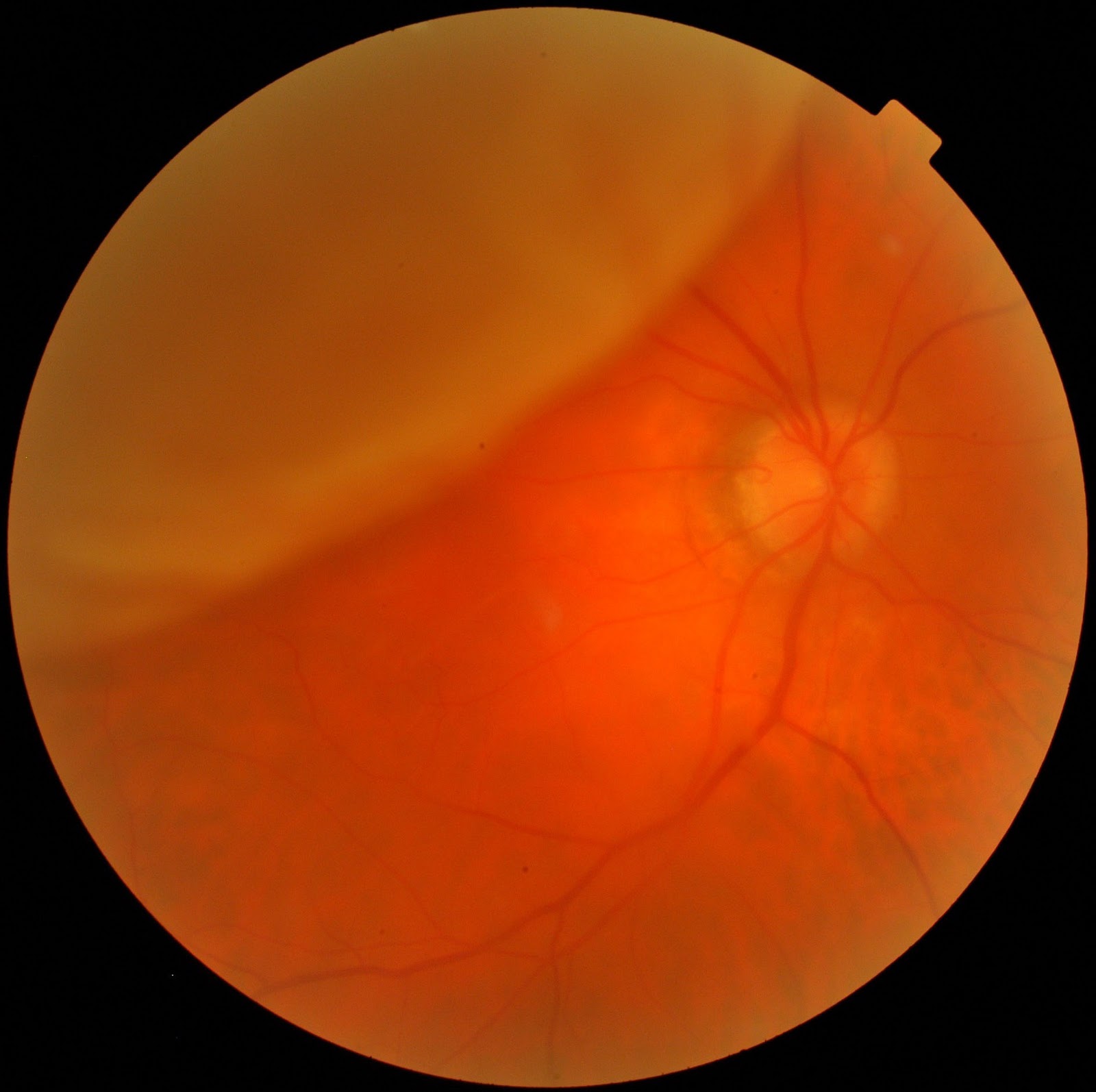
A 45-year-old phakic man has a macula-on detachment with one horseshoe tear at 11 o’clock and a clear view.
Pneumatic retinopexy is also offered, with laser and a gas bubble, and he is positioned face-down for a few days (see macula-off retinal detachment for the urgency of macula-threatening cases).
The retina flattens, and he keeps 20/20 vision, with no cataract surgery, which shows how a simple method can fit the right case.
Urgency and Timing
Macula-on detachments need prompt repair, ideally within a day or two, and macula-off detachments within a few days, since the outcome of the macula depends on the duration of detachment.
Until surgery, patients should limit activity and keep a head position that helps the retina to settle.
Reoperations and Proliferative Disease
About one in ten eyes need another operation, and proliferative vitreoretinopathy is the main reason (see methotrexate for proliferative vitreoretinopathy).
Silicone oil is used in complex cases and removed when the retina is stable (see silicone oil removal).
Modern Retinal Detachment Repair: Follow-Up After Repair
- Review at one day, one week, and one month, with a dilated peripheral examination
- Check the pressure, since gas can raise it
- Remind patients not to fly or go to altitude with gas in the eye (see intraocular gas and air travel)
- Examine the fellow eye and treat risky lesions if present (see fellow eye retinal detachment risk)
- Plan cataract surgery after vitrectomy when needed
Prevention and Early Detection
Examine patients with new flashes and floaters within days, since treating a tear with laser can prevent a detachment (see retinal detachment symptoms and triage).
In addition, education about warning signs is one of the most effective ways to improve outcomes, and it costs very little.
High-risk groups, such as highly myopic patients and those with a detachment in the other eye, should be told to report symptoms at once.


Document what you see
Two smartphone imaging tools built for everyday clinic use — one for the slit lamp, one for the fundus.
From Choroida — the team behind this siteReferences
- Hillier RJ, Felfeli T, Berger AR, et al. The Pneumatic Retinopexy versus Vitrectomy for the Management of Primary Rhegmatogenous Retinal Detachment Outcomes Randomized Trial (PIVOT). Ophthalmology. 2019;126:531-539.
- Heimann H, Bartz-Schmidt KU, Bornfeld N, et al. Scleral buckling versus primary vitrectomy in rhegmatogenous retinal detachment: a prospective randomized multicenter clinical study. Ophthalmology. 2007;114:2142-2154.
- Ryan SJ, Schachat AP, Wilkinson CP, et al, eds. Retina. 6th ed. Elsevier; 2018.
- American Academy of Ophthalmology. Basic and Clinical Science Course, Section 12: Retina and Vitreous. AAO; current edition.

Writes and reviews the clinical articles on Educate. Every post is checked against peer-reviewed sources before it goes live.