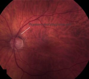
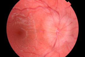
A normal retinal arteriole shows a thin, bright central light reflex, about a quarter to a third of the vessel width.
Over years of hypertension the wall thickens with hyaline material and the reflex changes.
Describing it as copper or silver wire is a quick, useful shorthand for how advanced the sclerosis is.
Copper wiring
The central reflex widens and turns a coppery, brownish-orange color.
The blood column is still just visible through the thickened wall.
It represents moderate arteriolosclerosis and is seen in the middle stages of long-standing hypertension.
Silver wiring
The wall becomes so opaque that the blood column cannot be seen, and the entire arteriole looks like a silver thread.
This implies advanced sclerosis, and in many cases the lumen has been seriously narrowed or obliterated.
In the extreme, the vessel becomes a whitish cord, sometimes called a “ghost” vessel.
Fundus Explorer Pro
Photograph the retinal findings described here with the phone already in your pocket — 22 D optics and built-in illumination in one handheld unit.
From Choroida — the team behind this siteWhat they reflect
The light reflex is determined by wall thickness and transparency.
These signs therefore tell you about chronicity and vessel wall status, not about current blood pressure.
A patient with silver wiring has had a long exposure to hypertension, atherosclerosis, or both, and a control reading on the day of the visit may look entirely normal.
Associated findings
- AV crossing changes (see arteriovenous crossing signs)
- Generalized arteriolar narrowing
- Retinal vein occlusions, where a hardened arteriole is the culprit (see central retinal vein occlusion)
- Retinal arterial macroaneurysm
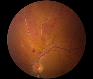
- In malignant hypertension, cotton wool spots, hemorrhages and disc swelling superimposed on the chronic picture (see hypertensive choroidopathy)
How the reflex is produced
The central streak is light reflected from the interface between the blood column and the vessel wall.
When the wall is thin and transparent, you see a narrow bright line from the blood column itself.
As the wall thickens, light is scattered by the wall, the streak broadens, and the color of the underlying blood is lost, which is why copper (a mix of wall reflection and blood color) precedes silver (wall reflection only).
Diagnostic accuracy is modest.
Studies comparing observers show that agreement is moderate at best, and wire signs are no longer central to modern hypertensive retinopathy classification.
They remain useful because they can be recognized without a camera, in any clinic with an ophthalmoscope.
Related changes in the retinal vessels
- Arteriolar narrowing, assessed as a reduced arteriole to vein ratio (normally about two thirds)
- Focal constriction, where isolated segments narrow, usually in younger patients with acute rises in pressure
- Perivascular sheathing in long-standing disease, often confused with vasculitis (see retinal vasculitis sheathing patterns)
- Tortuosity and loss of the normal branching angle
Pitfalls
Wire signs are subjective, and they depend on the camera and the pigmentation of the fundus.
A heavily pigmented fundus can make a normal reflex appear broad.
Compare both eyes and examine several vessels before concluding anything.
Management and prognosis
There is no ocular therapy for the vessel wall.
Management means a thorough cardiovascular assessment, and, where relevant, imaging or specialist referral.
Retinal signs predict stroke and coronary events independently of office blood pressure, which is why it is worth documenting them in the chart.
What the signs do not tell you
Wire changes reflect the state of the wall. They do not measure the current blood pressure, and they do not distinguish hypertensive sclerosis from the sclerosis of ageing, diabetes, or atherosclerosis. A patient with silver wiring may have well-controlled pressure today, and a patient with an acute hypertensive crisis may show no wire changes at all, because acute pressure rises produce focal narrowing, hemorrhages, and cotton wool spots instead.
Using the finding in a consultation
Treat wire changes as a prompt to ask about long-standing hypertension, smoking, diabetes, and family history, and to look at the rest of the fundus for evidence of end-organ damage. Cotton wool spots, flame hemorrhages, and disc swelling suggest accelerated disease and need urgent medical attention. If the signs are isolated and the blood pressure is normal in clinic, a letter to the primary physician with a request for ambulatory monitoring is a reasonable step.
Photographs taken at regular intervals document whether the changes progress, which is more informative than a single grading.
A short case to illustrate
A 58-year-old man attends for a routine eye test. He feels well and has not seen a doctor for years. The fundus shows broad, coppery light reflexes on most arterioles, several crossings with nicking, and one old branch vein occlusion with sheathed vessels. His clinic blood pressure is high. The retinal picture tells you that the hypertension has been present for a long time, which changes the conversation from a single high reading to a chronic problem that needs investigation and long-term treatment.
Such a finding is a good reason to write to the primary physician, because ophthalmologists are sometimes the first to detect the consequences of undiagnosed hypertension.


Document what you see
Two smartphone imaging tools built for everyday clinic use — one for the slit lamp, one for the fundus.
From Choroida — the team behind this siteReferences
- Keith NM, Wagener HP, Barker NW. Some different types of essential hypertension: their course and prognosis. Am J Med Sci. 1939;197:332-343.
- Walsh JB. Hypertensive retinopathy. Description, classification, and prognosis. Ophthalmology. 1982;89:1127-1131.
- Wong TY, Klein R, Couper DJ, et al. Retinal microvascular abnormalities and incident stroke: the Atherosclerosis Risk in Communities Study. Lancet. 2001;358:1134-1140.
- Wong TY, Mitchell P. The eye in hypertension. Lancet. 2007;369:425-435.
Test yourself
A few questions straight from this article.
-
How wide is the central light reflex on a normal retinal arteriole?
A normal arteriole shows a thin bright central reflex occupying roughly a quarter to a third of the vessel width; widening of that streak is the first change of sclerosis. -
What does copper wiring of a retinal arteriole signify?
In copper wiring the reflex widens and turns brownish-orange while the blood column remains just visible, reflecting moderate sclerosis in the middle stages of long-standing hypertension. -
What distinguishes silver wiring from copper wiring of a retinal arteriole?
In silver wiring the wall is opaque enough to hide the blood column and the whole arteriole looks like a silver thread, implying advanced sclerosis with a narrowed or obliterated lumen. -
What determines the appearance of the arteriolar light reflex?
The reflex is determined by wall thickness and transparency, which is why these signs report on vessel wall status and chronicity rather than on the current blood pressure. -
A hardened retinal arteriole is held responsible for which associated complication?
Retinal vein occlusion is listed among the associated findings, the hardened arteriole being the culprit at the site of compression. -
Why does copper wiring appear before silver wiring as sclerosis advances?
Copper is a mix of wall reflection and the colour of the underlying blood; once the wall scatters all the light and the blood colour is lost, only wall reflection remains, giving silver. -
Which fundus changes does an acute hypertensive crisis produce rather than wire changes?
Acute pressure rises produce focal narrowing, haemorrhages and cotton wool spots, so a patient in crisis may show no wire changes at all. -
What is the normal retinal arteriole to vein ratio used when judging arteriolar narrowing?
Arteriolar narrowing is assessed as a reduced arteriole to vein ratio, which is normally about two thirds. -
Which pitfall limits the reliability of wire signs on fundus examination?
Wire signs are subjective and depend on the camera and fundus pigmentation, so several vessels and both eyes should be compared before drawing a conclusion. -
What is the clinical value of documenting copper or silver wiring?
There is no ocular therapy for the vessel wall; the value lies in prompting cardiovascular assessment, since retinal signs predict stroke and coronary events independently of office blood pressure.

Writes and reviews the clinical articles on Educate. Every post is checked against peer-reviewed sources before it goes live.