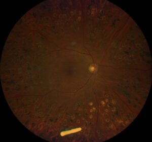
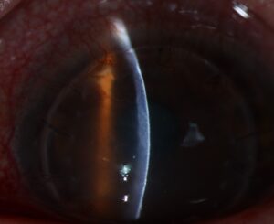
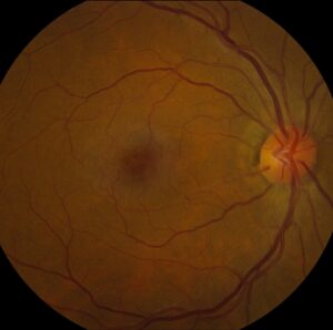
Retinal detachment in high myopia is a common cause of rhegmatogenous detachment, and it shows different features from detachment in eyes of normal axial length.
Longer eyes have thinner retina, more peripheral degeneration, and an earlier posterior vitreous detachment, and the combination raises the lifetime risk several-fold.
The surgeon must plan for atypical breaks, a macular hole in some patients, and a higher chance of redetachment.
Retinal Detachment in High Myopia: Why the Risk Rises
Axial elongation stretches the retina and the sclera, and the peripheral retina becomes thinner and more prone to degenerative change.
Liquefaction of the vitreous begins earlier in myopic eyes, and a posterior vitreous detachment often develops a decade or more earlier than in emmetropes (see vitreous syneresis).
Lattice degeneration is more frequent in myopia, and it is strongly associated with atrophic holes and tears at its edges (see lattice degeneration).
Eyes with an axial length above about 26 millimeters have a risk of detachment that is many times higher than that of emmetropic eyes, although absolute numbers remain small.
Cataract surgery, particularly with posterior capsule rupture and vitreous loss, adds to the risk in myopic eyes, as does clear lens extraction.
Clinical Features
- Detachments in younger patients than in the general population
- Multiple small atrophic holes, often within lattice, and sometimes a very peripheral dialysis
- A slow, shallow detachment with few symptoms, because atrophic holes release little traction
- Macular hole retinal detachment in eyes with posterior staphyloma, in which the detachment begins at the posterior pole
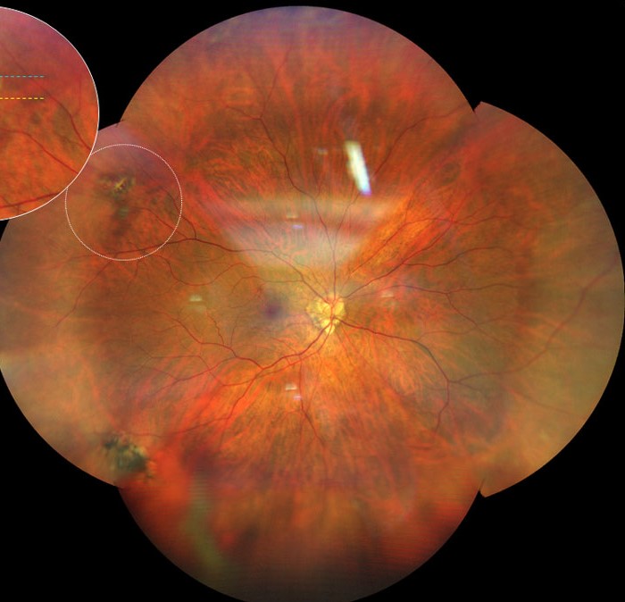
- Extensive pigmentation at the edge of an old, demarcated detachment line
Posterior staphyloma and chorioretinal atrophy make the retina difficult to examine, and the posterior pole may be tilted and hard to view.
Fundus Explorer Pro
Photograph the retinal findings described here with the phone already in your pocket — 22 D optics and built-in illumination in one handheld unit.
From Choroida — the team behind this siteMacular Hole Retinal Detachment
In eyes with pathologic myopia, a full-thickness macular hole may lead to a posterior pole detachment, with the fluid confined to the macula and arcades.
The mechanism includes traction from a thin posterior hyaloid, epiretinal membrane, and the staphyloma itself, which stretches the retina.
Pars plana vitrectomy with careful removal of the posterior cortex and internal limiting membrane, gas or silicone tamponade, and sometimes inverted flap techniques are used.
Macular buckling is an alternative that supports the posterior wall of the eye (see macular buckle surgery).
Evaluation
Perform dilated indirect ophthalmoscopy with scleral depression in every high myope with floaters or flashes, and examine the other eye as well.
Widefield imaging and OCT of the posterior pole help to find hidden breaks and to assess the staphyloma.
B-scan ultrasonography helps when the view is poor or the cornea or lens is not clear.
Treat symptomatic breaks and lattice with associated tears using laser retinopexy.
Prophylactic treatment of asymptomatic lattice without breaks is not routine, because evidence of benefit is limited.
Surgical Options
The choice depends on the type and location of breaks, the lens status, and the posterior pole.
- Scleral buckle: often favored in young myopes with attached vitreous and no posterior vitreous detachment (see scleral buckle surgery)
- Pars plana vitrectomy with gas: useful with posterior breaks, a macular hole, or traction (see pars plana vitrectomy)
- Combined procedures: for complex detachments with proliferative vitreoretinopathy
Redetachment is more frequent in myopic eyes, and the patient should know this in advance.
Counselling includes advice about the symptoms of recurrence and regular follow-up, because the fellow eye also needs surveillance.
Prophylaxis and the Fellow Eye
The fellow eye of a myope with detachment has a higher chance of breaks, and a careful examination with scleral depression is standard.
Laser or cryotherapy for symptomatic horseshoe tears is accepted practice, and many surgeons also treat lattice with holes in an eye whose fellow has detached.
For asymptomatic lattice alone, the evidence for treatment is weak, and the decision should be made together with the patient.
Counselling and Follow-Up
Patients with high myopia should know that their risk is higher than average, and that new floaters, flashes, or a shadow need a prompt visit.
Advise caution with contact sports, and discuss the risk before any intraocular surgery, including clear lens extraction.
After a repair, follow-up includes surveillance for redetachment, macular changes, and the development of cataract.
Myopic patients also need regular review for glaucoma and macular disease, so a single visit can cover several risks.
Document the axial length, the refractive error, and any staphyloma in the chart at the first visit, because these numbers help to judge the risk and to plan any future intraocular surgery in the same eye.
Share the plan with the patient’s optometrist so that new symptoms reach the retina clinic quickly.


Document what you see
Two smartphone imaging tools built for everyday clinic use — one for the slit lamp, one for the fundus.
From Choroida — the team behind this siteReferences
- Ripandelli G, Coppé AM, Parisi V, et al. Posterior vitreous detachment and retinal detachment after cataract surgery. Ophthalmology. 2007;114:692-697.
- Ohno-Matsui K, Lai TY, Lai CC, Cheung CM. Updates of pathologic myopia. Prog Retin Eye Res. 2016;52:156-187.
- Ryan SJ, Schachat AP, Wilkinson CP, et al, eds. Retina. 6th ed. Elsevier; 2018.
- American Academy of Ophthalmology. Basic and Clinical Science Course, Section 12: Retina and Vitreous. AAO; current edition.
Test yourself
A few questions straight from this article.
-
Above approximately what axial length does retinal detachment risk rise many-fold in high myopia?
Eyes longer than about 26 millimeters carry a detachment risk many times that of emmetropic eyes, though the absolute numbers stay small. -
In myopic eyes, when does posterior vitreous detachment develop compared with emmetropic eyes?
Vitreous liquefaction starts earlier in myopia, so posterior vitreous detachment often arrives a decade or more before it would in an emmetrope. -
Why are atrophic-hole detachments in high myopia often shallow, slow and nearly symptomless?
Atrophic holes release very little traction, so fluid accumulates slowly and the detachment can stay shallow with few symptoms. -
In myopic macular hole retinal detachment, where does the subretinal fluid typically stay confined?
A full-thickness macular hole in pathologic myopia produces a posterior pole detachment, with fluid limited to the macula and the arcades. -
Which combination of forces drives macular hole retinal detachment in pathologic myopia?
Traction from a thin posterior hyaloid and epiretinal membrane combines with stretching by the staphyloma itself. -
Which step is central to vitrectomy for myopic macular hole retinal detachment?
Careful removal of the posterior cortex and the internal limiting membrane, with gas or silicone tamponade, is the core of the repair. -
In which high-myopic detachment is scleral buckle often the favored procedure?
Buckling suits young myopic eyes whose vitreous is still attached; posterior breaks, macular holes or traction push the choice toward vitrectomy. -
What is the accepted stance on treating asymptomatic lattice degeneration without breaks in high myopia?
Prophylactic treatment of asymptomatic lattice without breaks is not routine; the evidence of benefit is limited and the decision is shared with the patient. -
Which test helps assess a suspected myopic detachment when the fundus view is poor?
B-scan ultrasonography is the fallback when media opacity or a hazy cornea or lens prevents an adequate view of the retina. -
What should a high myope be warned about regarding the outcome of detachment repair?
Myopic eyes redetach more often, so patients are counselled in advance about recurrence symptoms and the need for continued follow-up of both eyes.

Writes and reviews the clinical articles on Educate. Every post is checked against peer-reviewed sources before it goes live.