
Scleral buckle surgery is an extraocular technique for repairing rhegmatogenous retinal detachment by indenting the sclera from the outside to bring the pigment epithelium back into contact with the detached retina.
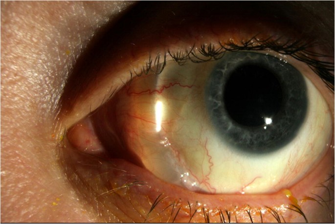
It predates vitrectomy by decades and remains a first-line option for a defined subset of detachments, particularly in phakic eyes with well-localized peripheral pathology.
Understanding when to reach for a buckle rather than a vitrectomy, and what the procedure actually does mechanically, still matters even in a vitrectomy-dominant era.
The technique traces back to the mid-twentieth-century work of Custodis and later Schepens, whose refinements established the principle that closing a retinal break from the outside, without ever entering the eye, could reliably reattach the retina.
What the Buckle Actually Does
A scleral buckle is a silicone band, sponge, or solid element sutured onto the outer scleral surface, indenting the eye wall inward toward the vitreous cavity.
This indentation relieves vitreoretinal traction at the site of a retinal break by moving the retinal pigment epithelium closer to the retina rather than by removing vitreous or gas-tamponading the break from inside, which is the vitrectomy approach.
The buckle is typically left in place permanently, functioning passively for the rest of the eye’s life rather than being removed once the retina has settled.
Indications
Scleral buckling is best suited to phakic eyes with a single break or a cluster of breaks confined to one quadrant, relatively fresh detachments without significant proliferative vitreoretinopathy, and eyes with good vitreous support where a pneumatic or laser-only approach is not appropriate.
- Young phakic patients, where avoiding vitrectomy-associated cataract progression is a meaningful consideration
- Detachments with breaks anterior to the equator, which are easier to support externally than to treat from inside
- Eyes with clear media, allowing accurate intraoperative localization of every break under indirect ophthalmoscopy
- Combined scleral buckle plus vitrectomy, used when there is a mix of anterior and posterior pathology that neither approach alone would adequately address
Pseudophakic and posterior-pole detachments have shifted heavily toward primary vitrectomy over the past two decades, since visualizing and treating posterior breaks externally is far less reliable than doing so internally.
Fundus Explorer Pro
Photograph the retinal findings described here with the phone already in your pocket — 22 D optics and built-in illumination in one handheld unit.
From Choroida — the team behind this siteSurgical Technique
The surgeon opens the conjunctiva, isolates the rectus muscles, and searches the entire retinal periphery with scleral depression and indirect ophthalmoscopy to localize every break, since missing even one undermines the whole repair.
Cryotherapy or, less commonly, laser is applied around each break to create a chorioretinal adhesion, and the buckle material is then sutured over the marked area to achieve the calculated indentation height and width.
Subretinal fluid may be drained externally through a small sclerotomy if the detachment is bullous enough that the buckle alone would not appose the retina promptly, though many surgeons now avoid drainage when possible given its own complication profile.
An encircling band is added in eyes with multiple breaks spread across more than one quadrant, or with diffuse peripheral pathology such as lattice degeneration, to support the entire 360 degrees of retina rather than just the immediate area of the break.
Buckle material choice matters clinically: a soft silicone sponge segment is often used for a localized break, while a firmer solid silicone tire is favored when broader, more uniform indentation is needed under an encircling band.
Comparison With Vitrectomy
The SPR study, a prospective randomized multicenter trial comparing the two approaches, found broadly comparable anatomic and functional outcomes in phakic eyes, with a modest myopic shift and higher initial reoperation rate in the buckle group but a lower rate of cataract progression than in the vitrectomy group.
This trial and the literature that followed it are the basis for the current, fairly durable consensus: buckle and vitrectomy are not competing universally, they are matched to different anatomic scenarios, and the choice should be individualized rather than defaulting reflexively to vitrectomy for every case.
Complications
- Induced myopia from the added axial length of the indentation, usually 1-3 diopters and largely predictable from buckle height
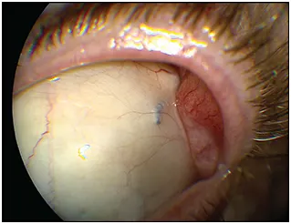
- Diplopia from rectus muscle disinsertion and reattachment during exposure, usually transient but occasionally persistent
- Buckle infection or extrusion, which can present months to years postoperatively and sometimes requires removal
- Anterior segment ischemia, a rare but serious complication most associated with extensive encircling procedures combined with vortex vein compromise
- Recurrent detachment from a new break, missed break, or proliferative vitreoretinopathy developing after apparently successful surgery
- Persistent strabismus from scarring or slippage of a rectus muscle, which occasionally needs its own dedicated correction once the retinal repair has stabilized
Postoperative Course
Patients are followed closely in the first postoperative weeks to confirm the retina has settled and to catch any new break early, since the peripheral retina remains at some ongoing risk from the same predisposing factors that caused the original detachment.
The buckle itself is not routinely removed unless it becomes symptomatic, infected, or is thought to be contributing to a later complication, and most patients tolerate a well-placed buckle indefinitely without ever needing a second procedure directed at the implant itself.
Visual recovery depends heavily on whether the macula was detached before surgery and for how long, which is why the same urgency that applies to any rhegmatogenous detachment applies here regardless of which surgical technique is ultimately chosen.


Document what you see
Two smartphone imaging tools built for everyday clinic use — one for the slit lamp, one for the fundus.
From Choroida — the team behind this siteReferences
- Heimann H, Bartz-Schmidt KU, Bornfeld N, et al. Scleral buckling versus primary vitrectomy in rhegmatogenous retinal detachment: a prospective randomized multicenter clinical study. Ophthalmology.
- Schwartz SG, Flynn HW Jr. Primary retinal detachment: scleral buckle or pars plana vitrectomy? Current Opinion in Ophthalmology.
- American Academy of Ophthalmology. Basic and Clinical Science Course, Section 12: Retina and Vitreous.
- Wilkinson CP, Rice TA. Michels’ Retinal Detachment.
Test yourself
A few questions straight from this article.
-
How does a scleral buckle reattach the retina in rhegmatogenous retinal detachment?
The buckle is sutured to the outer sclera and indents the eye wall inward, relieving vitreoretinal traction at the break by moving the pigment epithelium back into contact with the detached retina. -
Which eye is best suited to primary scleral buckling?
Buckling suits phakic eyes with a single break or cluster of breaks in one quadrant, relatively fresh detachments without significant proliferative vitreoretinopathy, and clear media. -
Why have posterior-pole and pseudophakic detachments shifted toward primary vitrectomy?
Visualising and treating posterior breaks from outside the eye is far less reliable than doing so internally, which is why these cases have moved toward primary vitrectomy. -
How does the surgeon localise every retinal break during scleral buckle surgery?
The entire retinal periphery is searched with scleral depression and indirect ophthalmoscopy, because missing even a single break undermines the whole repair. -
What creates the chorioretinal adhesion around each break in scleral buckle surgery?
Cryotherapy, or less commonly laser, is applied around each break to create a chorioretinal adhesion before the buckle is sutured over the marked area. -
When is an encircling band added during scleral buckle surgery?
An encircling band supports the full 360 degrees of retina and is added for breaks spread across more than one quadrant or diffuse peripheral pathology such as lattice degeneration. -
In the SPR trial, which outcome favoured scleral buckling over vitrectomy in phakic eyes?
The SPR trial found broadly comparable anatomic and functional outcomes in phakic eyes, with a modest myopic shift and higher initial reoperation rate after buckling, but less cataract progression than after vitrectomy. -
How much induced myopia does a scleral buckle typically produce?
The added axial length from the indentation usually induces 1 to 3 dioptres of myopia, an amount largely predictable from the buckle height. -
Which situation carries the greatest risk of anterior segment ischaemia after scleral buckling?
Anterior segment ischaemia is rare but serious, and is most associated with extensive encircling procedures combined with vortex vein compromise. -
What most influences visual recovery after scleral buckle surgery?
Visual recovery depends heavily on whether the macula was detached before surgery and for how long, which is why the usual urgency applies whichever technique is chosen.
