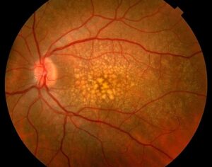
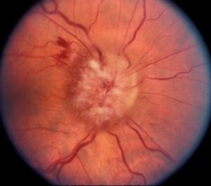
Vitreopapillary traction (VPT) occurs when the vitreous remains attached to the optic disc during a posterior vitreous detachment and pulls on it.
It can also cause disc swelling, hemorrhage, and retinal edema.
It is one of the benign conditions that can look like disc swelling from raised intracranial pressure.
OCT usually settles the diagnosis in seconds.
What Is Vitreopapillary Traction?
In a posterior vitreous detachment, the vitreous gel separates from the retina.
The last place to let go is often the optic disc, where the adhesion is strong (see posterior vitreous detachment).
If the separation is incomplete, the residual adhesion pulls on the disc and the surrounding retina.
The traction may also be tangential or anteroposterior.
It can be present alone or with vitreomacular traction (see vitreomacular traction).
Sebag has described anomalous detachment as a mechanism that causes such vitreoretinal interface disease (see vitreoschisis).
Clinical Features of Vitreopapillary Traction
- Mild to moderate blurring of vision, or often no symptoms
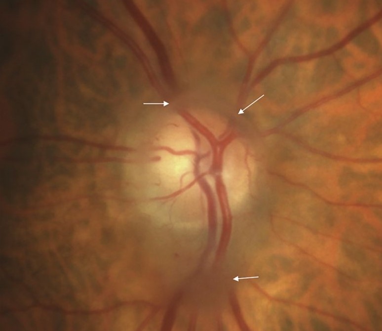
- Unilateral disc swelling or elevation, usually without a relative afferent pupillary defect
- Peripapillary or intrapapillary hemorrhages, sometimes striking
- Peripapillary retinal edema, retinoschisis, or fluid
- Floaters and flashes in some patients, due to the vitreous detachment
- Visual field defects that are minimal or related to the blind spot
The patient is typically middle-aged or older, and the myopic are over-represented.
Young patients with an incomplete detachment can also be affected, with disc and peripapillary hemorrhage described in that group (see the references).
Fundus Explorer Pro
Photograph the retinal findings described here with the phone already in your pocket — 22 D optics and built-in illumination in one handheld unit.
From Choroida — the team behind this siteWhy It Can Be Mistaken for Papilledema
- The disc looks swollen and elevated, with blurred margins
- Hemorrhages may be present
- The patient may also have headache or visual symptoms for unrelated reasons
Several features suggest traction and not a systemic cause.
- Unilateral involvement
- No venous engorgement or loss of spontaneous venous pulsations in a manner consistent with raised pressure
- Evidence of a partial vitreous detachment
OCT Findings
OCT of the disc shows the posterior hyaloid as a thin line, attached to the disc and elevated elsewhere, with a tent-like traction on the peripapillary retina.
Also, peripapillary retinoschisis or fluid and thickening of the retinal nerve fiber layer may be seen.
The scan after the release of traction shows resolution, and the swelling often settles within weeks to months.
In addition, widefield and swept-source imaging can show the extent of the detachment (see OCT retinal layers).
Differential Diagnosis of Vitreopapillary Traction
- Papilledema from raised intracranial pressure: bilateral disc swelling with venous changes and field enlargement. A need for systemic evaluation (see papilledema)
- Optic disc drusen: a crowded disc with buried drusen that can also be seen on OCT (see optic disc drusen)
- Anterior ischemic optic neuropathy and optic neuritis (see optic neuritis)
- Papillophlebitis and retinal vein occlusion (see papillophlebitis)
- Diabetic papillopathy and infiltrative disease
Management for Vitreopapillary Traction
Most cases resolve by themselves when the vitreous separates fully, and they need no treatment.
- First, observe with OCT at intervals of a few weeks
- Examine the periphery for retinal tears, since the detachment can cause breaks (see horseshoe tears)
- Also inform the patient about the warning signs of retinal detachment
- Consider vitrectomy if traction persists with vision loss or macular involvement
Urgent neuroimaging is not usually needed if the picture is classic and the OCT shows traction, but any atypical feature should prompt further evaluation.
Counselling
Explain that the swelling is caused by the gel in the eye pulling on the disc, that it usually settles by itself, and that a few follow-up visits will confirm this.
Also, reassure the patient that the finding does not usually indicate a serious disease of the brain.
Documenting and Following the Case
Record the OCT findings, the visual acuity, and the visual field at the first visit, and repeat the scan at intervals of two to four weeks until the traction has resolved.
A change in the findings, such as a growing area of fluid or new hemorrhages, calls for a closer look at the periphery and macula.
When to Consider Further Tests
If the disc swelling is bilateral, if there is headache with visual obscurations, if the acuity or the field is out of proportion, or if the OCT does not show traction, other causes should be pursued.
In these cases, a neuro-ophthalmic evaluation and imaging may also be needed, according to the findings.
A Typical Case
A man in his late fifties notices floaters, and his optometrist finds a swollen right disc with a few flame hemorrhages.
OCT shows a hyaloid line pulling on the disc, with peripapillary fluid, and the left disc is normal.
The acuity is 20/20, the field is normal, and the pupils are equal and reactive.
The periphery is examined and shows no breaks, and follow-up scans over six weeks show release of traction and resolution of the swelling.


Document what you see
Two smartphone imaging tools built for everyday clinic use — one for the slit lamp, one for the fundus.
From Choroida — the team behind this siteReferences
- Katz B, Hoyt WF. Intrapapillary and peripapillary hemorrhage in young patients with incomplete posterior vitreous detachment: signs of vitreopapillary traction. Ophthalmology. 1995;102:349-354.
- Sebag J. Anomalous posterior vitreous detachment: a unifying concept in vitreo-retinal disease. Graefes Arch Clin Exp Ophthalmol. 2004;242:690-698.
- Ryan SJ, Schachat AP, Wilkinson CP, et al, eds. Retina. 6th ed. Elsevier; 2018.
- American Academy of Ophthalmology. Basic and Clinical Science Course, Section 12: Retina and Vitreous. AAO; current edition.

Writes and reviews the clinical articles on Educate. Every post is checked against peer-reviewed sources before it goes live.