The choriocapillaris is the thin capillary layer of the choroid that feeds the outer retina.
It lies directly beneath Bruch’s membrane and the retinal pigment epithelium.
Therefore, damage to it harms photoreceptors long before other signs appear.
Position and Structure
The choriocapillaris is a single layer of wide, fenestrated capillaries about 10 micrometers thick.
It sits between Bruch’s membrane and the medium vessels of Sattler’s layer.
Also, the fenestrations face the pigment epithelium.
As a result, nutrients and oxygen cross easily to the outer retina.
Lobular Organization
The capillaries form hexagonal lobules fed by a feeder arteriole and drained by venules.
Lobules are small in the posterior pole and elongated in the periphery.
Also, neighboring lobules share little flow.
Therefore, loss of a feeder arteriole can silence a whole unit.
Fundus Explorer Pro
Photograph the retinal findings described here with the phone already in your pocket — 22 D optics and built-in illumination in one handheld unit.
From Choroida — the team behind this siteBlood Flow of the Choriocapillaris
The choroid has the highest blood flow per tissue weight of any tissue in the body.
This high flow carries heat and oxygen away from the fovea.
Also, the low oxygen extraction keeps the venous blood bright red.
As a result, small drops in flow can still support the retina for a while.
Choriocapillaris and the Retinal Pigment Epithelium
The pigment epithelium depends on the capillary layer for glucose and oxygen.
In turn, it sends VEGF to the capillaries to keep them open.
Also, loss of one partner leads to loss of the other.
Therefore, the two form a single functional unit.
Imaging the Choriocapillaris with OCT Angiography
OCT angiography shows flow in the choriocapillaris as a fine granular pattern.
Dark areas without signal are called flow deficits.
Also, the thin layer sits under the pigment epithelium, so shadowing can mimic deficits.
Therefore, interpret flow deficits with care.
Artifacts and Pitfalls in Imaging
Drusen, pigment epithelial detachments and media opacity create false deficits.
Projection from the retinal vessels can add false signal.
Also, the slab thickness and segmentation errors change the result.
As a result, check the structural scan for each deficit.
Flow Deficits in Aging and AMD
Flow deficit density rises with age.
Mullins and colleagues showed capillary loss around drusen in early disease.
Also, eyes with geographic atrophy show large deficits beyond the edge of atrophy.
Therefore, capillary loss may precede the death of the pigment epithelium.
Choriocapillaris in Diabetes and Hypertension
Diabetic eyes show flow deficits even without retinopathy.
Hypertensive choroidopathy produces patchy nonperfusion of lobules (see hypertensive choroidopathy).
Also, eyes with preeclampsia show similar findings.
In addition, the changes can reverse with control.
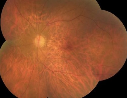
Choriocapillaris in Inflammation
Choroiditis and punctate inner choroidopathy cause areas of nonperfusion.
Acute posterior multifocal placoid pigment epitheliopathy shows large deficits.
Also, deficits can resolve with treatment.
As a result, OCT angiography helps to track disease activity.
Choriocapillaris in Genetic Disease
Pseudoxanthoma elasticum and Sorsby fundus dystrophy damage the layer.
Choroideremia leads to atrophy of the capillaries with the epithelium.
Also, Stargardt disease may show deficits around flecks.
Therefore, inherited diseases can start in the vascular layer.
Measuring Choriocapillaris Flow
Researchers count the percent of deficits and their size.
Spaide described a power law distribution of flow features that helps to compare eyes.
Also, the measures depend on the device and the scan size.
Therefore, compare only scans from the same system.
Relation to Choroidal Thickness
The capillary layer is a tiny part of the choroidal thickness.
Thick choroid and thin choriocapillaris occur together in pachychoroid disease.
Also, large vessels in the Haller layer can compress the layer above.
As a result, thickness alone does not predict capillary health.
Therapeutic Implications for the Choriocapillaris
Photodynamic therapy can cause temporary closure of the capillary layer.
Anti-VEGF therapy might thin the layer through loss of endothelial support.
Also, new complement inhibitors aim at the capillary damage seen in atrophy.
Therefore, capillary health matters to every treatment plan.
Clinical Use Today
OCT angiography of the choriocapillaris is a research tool in most clinics.
It helps in complex cases of unexplained outer retinal loss.
Also, repeated scans can show progression.
In addition, combine it with fluorescein and indocyanine angiography (see indocyanine green angiography).
Pitfalls to Avoid
- Do not call every dark area a flow deficit without checking artifacts
- Never compare scans from different devices
- Do not assume that thick choroid means healthy capillaries
- Never use one scan to judge progression
For the latest studies, see the PubMed results for choriocapillaris.


Document what you see
Two smartphone imaging tools built for everyday clinic use — one for the slit lamp, one for the fundus.
From Choroida — the team behind this siteReferences
- Nickla DL, Wallman J. The multifunctional choroid. Prog Retin Eye Res. 2010;29:144-168.
- Spaide RF. Choriocapillaris flow features follow a power law distribution: implications for characterization and mechanisms of disease progression. Am J Ophthalmol. 2016;170:58-67.
- Mullins RF, Johnson MN, Faidley EA, Skeie JM, Huang J. Choriocapillaris vascular dropout related to density of drusen in human eyes with early age-related macular degeneration. Invest Ophthalmol Vis Sci. 2011;52:1606-1612.
- Borrelli E, Sarraf D, Freund KB, Sadda SR. OCT angiography and evaluation of the choroid and choroidal vascular disorders. Prog Retin Eye Res. 2018;67:30-55.
Test yourself
A few questions straight from this article.
-
Where does the choriocapillaris lie?
The choriocapillaris sits directly beneath Bruch's membrane and the RPE, above the medium vessels of Sattler's layer. -
Approximately how thick is the choriocapillaris layer?
The choriocapillaris is a single layer of wide, fenestrated capillaries about 10 micrometers thick. -
Toward which structure do the fenestrations of the choriocapillaris face?
The fenestrations face the pigment epithelium, so nutrients and oxygen cross easily to the outer retina. -
How is the choriocapillaris organized into functional units?
The capillaries form hexagonal lobules fed by a feeder arteriole and drained by venules; neighbouring lobules share little flow, so losing a feeder arteriole can silence a whole unit. -
Which property characterizes choroidal blood flow?
The choroid has the highest blood flow per tissue weight of any tissue, and its low oxygen extraction keeps the venous blood bright red. -
Which signal from the retinal pigment epithelium keeps the choriocapillaris open?
The RPE depends on the choriocapillaris for glucose and oxygen and in turn sends VEGF to keep the capillaries open, so the two form one functional unit. -
Which findings can create false choriocapillaris flow deficits on OCT angiography?
Drusen, pigment epithelial detachments and media opacity create false deficits, whereas projection from retinal vessels adds false signal, so check the structural scan. -
In geographic atrophy, where do large choriocapillaris flow deficits extend?
Eyes with geographic atrophy show large deficits beyond the edge of atrophy, suggesting capillary loss may precede death of the pigment epithelium. -
When can choriocapillaris flow deficits appear in diabetic eyes?
Diabetic eyes show choriocapillaris flow deficits even without retinopathy. -
In pachychoroid disease, how do choroidal thickness and the choriocapillaris relate?
Thick choroid and thin choriocapillaris occur together in pachychoroid disease, as large Haller vessels can compress the layer above, so thickness alone does not predict capillary health.

Writes and reviews the clinical articles on Educate. Every post is checked against peer-reviewed sources before it goes live.