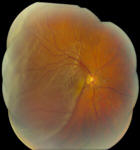
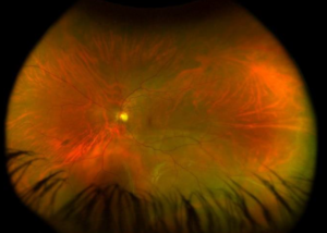
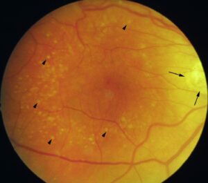
An intraocular foreign body (IOFB) is a fragment that has entered the eye and remains inside, and it is a form of open globe injury.
Also, hammering metal on metal is the classic mechanism, and the injury is often small, with the patient unaware of the seriousness.
Also, imaging, early repair, and measures against infection are the pillars of care.
Intraocular Foreign Body: Mechanism and Risk
High-speed fragments from hammering, grinding, and drilling can penetrate the cornea or sclera and lodge in the lens, vitreous, or retina.
In addition, other sources include gunshot pellets, wood and glass fragments, and stone.
The risks are also infection, damage from the entry and path, retinal tears and detachment, and toxic effects of the material.
Young men in industrial and agricultural jobs are also most affected, and eye protection is the main prevention.
Intraocular Foreign Body: Clinical Evaluation
- Take a careful history of the mechanism, the activity, the possible material, and the use of protection
- Check the visual acuity and the pupils, and look for an entry wound in the cornea or sclera
- Look for signs of perforation: shallow chamber, Seidel positive, iris defect, hyphema, and lens opacity
- Avoid pressure on the globe, and place a shield
- Examine the posterior segment if the media allow, and the other eye
- Give systemic and topical antibiotics, and check the tetanus status
Clinicians may miss a small, self-sealing entry wound, so keep a high index of suspicion (see ruptured globe).
Fundus Explorer Pro
Photograph the retinal findings described here with the phone already in your pocket — 22 D optics and built-in illumination in one handheld unit.
From Choroida — the team behind this siteImaging in Intraocular Foreign Body
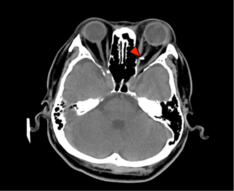
- CT of the orbits and brain with thin sections is the most sensitive test for metallic and many other foreign bodies, and it shows the location
- B-scan ultrasonography can show foreign bodies in the vitreous and detect retinal detachment, but it must be gentle in open globe injuries (see B-scan ultrasonography)
- Ultrasound biomicroscopy shows anterior segment fragments
- MRI is contraindicated if a magnetic metal is suspected, because the fragment can move and cause damage, but it is useful for wood and plastic when metal has been excluded
- Plain radiographs have limited sensitivity
Do not let imaging delay closure, and do the CT shortly after.
Management for Intraocular Foreign Body
Clinicians do primary closure of the entry wound promptly.
Surgeons usually remove the foreign body by pars plana vitrectomy within a few days in most cases, and sooner if there is infection or a reactive material such as copper.
- Magnetic fragments: removed with an intraocular magnet or forceps
- Nonmagnetic fragments: removed with forceps through a sclerotomy or the entry wound
- Anterior segment fragments: removed through a limbal incision
- Retinal tears and detachment: repaired with laser, buckle, or tamponade (see pars plana vitrectomy)
Surgeons may also leave some small inert fragments in the periphery in place, with follow-up.
Endophthalmitis Prophylaxis
Infection is a major risk, reported in several percent of cases, and it is higher with organic material and delayed closure.
In addition, prophylaxis includes systemic antibiotics, often a fluoroquinolone, and intravitreal antibiotics at the time of surgery in high-risk cases.
Take vitreous cultures if you suspect infection (see acute postoperative endophthalmitis).
Toxic Effects of Metals
- Siderosis bulbi: from retained iron, with retinal toxicity, pigment changes, lens deposits, and glaucoma (see siderosis bulbi)
- Chalcosis: from copper, which can cause severe inflammation and a Kayser-Fleischer-like ring, and requires early removal
- Other metals and organic materials: a variable reaction
The ERG can detect early retinal toxicity in siderosis.
Outcome and Follow-Up
The outcome depends on the initial acuity, the location and size of the fragment, infection, and retinal detachment.
Follow the patient for proliferative vitreoretinopathy, cataract, glaucoma, and late toxic effects.
Also, explain the risks to the patient, and advise protective eyewear at work.
Special Considerations
- Retained lens fragments and cataract: the surgeon may remove a fragment in the lens with cataract surgery if the posterior capsule is intact, and the lens status determines the approach
- Anterior chamber fragments: often removed early to avoid corneal damage and inflammation
- Fragments embedded in the retina: may be surrounded by laser before removal to reduce the risk of detachment
- Sympathetic ophthalmia: a rare late complication of penetrating injury that calls for follow-up for months (see sympathetic ophthalmia)
Occupational Prevention
Employers should provide goggles that meet safety standards, and the workers should wear them for hammering, grinding, and similar tasks.
After an injury, a report to the occupational health service and a review of the safety procedures help to prevent a second event.
In addition, remind patients that even a minor-looking entry wound needs to be examined, and that waiting increases the chance of infection.
Review patients at regular intervals for months after removal, with attention to inflammation, pressure, and the retina.
For the latest studies, see the PubMed results for intraocular foreign body.


Document what you see
Two smartphone imaging tools built for everyday clinic use — one for the slit lamp, one for the fundus.
From Choroida — the team behind this siteReferences
- Loporchio D, Mukkamala L, Gorukanti K, et al. Intraocular foreign bodies: a review. Surv Ophthalmol. 2016;61:582-596.
- Kuhn F, Morris R, Witherspoon CD, et al. A standardized classification of ocular trauma. Ophthalmology. 1996;103:240-243.
- Ryan SJ, Schachat AP, Wilkinson CP, et al, eds. Retina. 6th ed. Elsevier; 2018.
- American Academy of Ophthalmology. Basic and Clinical Science Course, Section 12: Retina and Vitreous. AAO; current edition.

Writes and reviews the clinical articles on Educate. Every post is checked against peer-reviewed sources before it goes live.