Normal-tension glaucoma is glaucomatous optic nerve damage and visual field loss occurring despite intraocular pressure that remains consistently within the statistically normal range, representing something close to the mirror image of ocular hypertension, discussed in its own dedicated article on this site.
If ocular hypertension is elevated pressure without damage, normal-tension glaucoma is damage without elevated pressure, and together these two conditions make clear that intraocular pressure, while central to glaucoma management, is neither necessary nor sufficient on its own to fully explain why a given optic nerve is being damaged.
Understanding this distinction reframes glaucoma more accurately as a disease of the optic nerve’s vulnerability relative to whatever pressure it experiences, rather than a disease defined purely by an absolute pressure threshold.
Normal-tension glaucoma accounts for a substantial proportion of open-angle glaucoma in some populations, particularly reported at higher relative rates in certain Asian populations compared to some Western cohorts, though it occurs worldwide.
Definition and Diagnostic Considerations
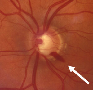
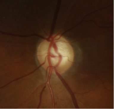
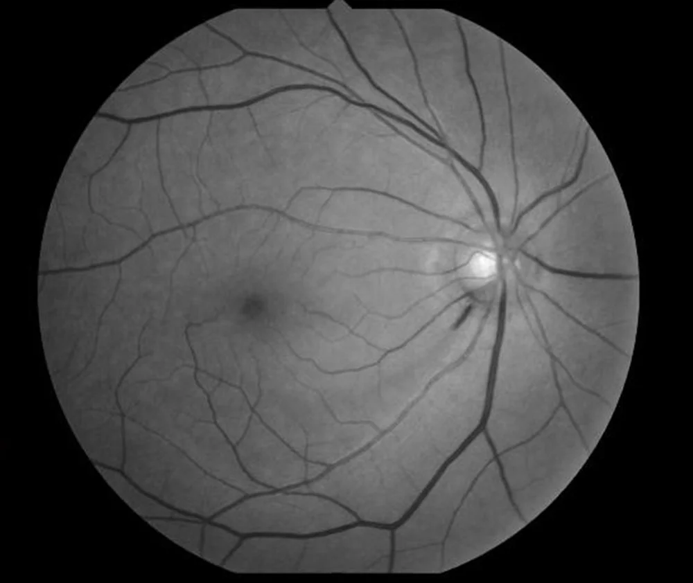
Normal-tension glaucoma is diagnosed when characteristic glaucomatous optic nerve damage and a corresponding visual field defect are present despite intraocular pressure measurements that consistently fall within the normal range on repeated testing.
Diurnal pressure variation needs careful consideration before accepting this diagnosis, since some patients have pressure spikes outside of typical office hours that a single daytime measurement would miss entirely, meaning a truly comprehensive pressure assessment, sometimes including measurements at different times of day, is warranted before concluding that pressure has genuinely remained normal throughout.
Other causes of optic nerve damage and field loss that can mimic glaucoma, including compressive lesions of the visual pathway, prior ischemic events, and other optic neuropathies, need to be actively excluded, since normal-tension glaucoma is, like several other conditions discussed on this site, partly a diagnosis of exclusion.
Neuroimaging is warranted when the field defect pattern, disc appearance, or overall clinical picture raises any doubt about a purely glaucomatous process, since missing a compressive lesion masquerading as normal-tension glaucoma would have serious consequences for the patient.
Proposed Mechanisms
Vascular dysregulation, an abnormal autoregulatory response of blood flow to the optic nerve head, is one of the leading proposed mechanisms, potentially leaving the nerve vulnerable to normal pressure levels that a nerve with typical vascular autoregulation would tolerate without damage.
Increased susceptibility of the optic nerve itself, whether from a structurally weaker lamina cribrosa or other individual anatomic variation, is another proposed contributor, suggesting some optic nerves are simply less tolerant of a given pressure level than others.
A low cerebrospinal fluid pressure in the space surrounding the optic nerve, creating a larger-than-usual pressure gradient across the lamina cribrosa even when intraocular pressure itself is normal, has also been proposed as a contributing mechanism in at least some patients.
All-fit Slit-Lamp Adapter
Record and share exactly what you see at the slit lamp. One adapter fits any slit lamp or surgical microscope — and any smartphone.
From Choroida — the team behind this siteClinical Associations
- Migraine and other vasospastic conditions, more commonly reported in patients with normal-tension glaucoma than in the general population, consistent with the vascular dysregulation hypothesis
- Systemic hypotension, including nocturnal blood pressure dipping, which some studies have associated with a higher risk of progression in normal-tension glaucoma specifically
- A visual field defect pattern that, in some series, tends to be more localized and closer to fixation at presentation compared to typical high-pressure glaucoma, though this pattern is not universal or diagnostically definitive on its own
Management
Lowering intraocular pressure further, even from an already statistically normal baseline, remains the primary evidence-based treatment strategy, since a major randomized trial demonstrated that reducing pressure by roughly 30 percent slowed progression in normal-tension glaucoma, mirroring the pressure-dependent treatment logic used in high-pressure glaucoma despite the different baseline pressure.
This finding is genuinely important: even though pressure was already “normal” before treatment, further lowering it still measurably slowed disease progression, reinforcing that pressure remains a meaningful, modifiable risk factor in this condition even when it does not appear elevated by standard population norms.
Addressing modifiable vascular risk factors, including avoiding significant nocturnal blood pressure dipping when medically appropriate to adjust, is considered alongside pressure-lowering treatment in managing patients where a vascular contribution seems plausible.
Standard topical, laser, and surgical glaucoma treatments, all discussed in their own dedicated articles on this site, are used to achieve the pressure reduction shown to be beneficial, following similar general principles to high-pressure glaucoma management.
Patients sometimes find a normal-tension glaucoma diagnosis conceptually confusing, since their pressure readings look reassuring on paper, and taking time to explain why treatment still helps despite an already normal pressure supports better long-term adherence to what can otherwise seem like a counterintuitive treatment plan.


Document what you see
Two smartphone imaging tools built for everyday clinic use — one for the slit lamp, one for the fundus.
From Choroida — the team behind this siteReferences
- Collaborative Normal-Tension Glaucoma Study Group. Comparison of glaucomatous progression between untreated patients with normal-tension glaucoma and patients with therapeutically reduced intraocular pressures. American Journal of Ophthalmology.
- American Academy of Ophthalmology. Basic and Clinical Science Course, Section 10: Glaucoma.
- Flammer J, Orgül S, Costa VP, et al. The impact of ocular blood flow in glaucoma. Progress in Retinal and Eye Research.
Test yourself
A few questions straight from this article.
-
Which combination defines normal-tension glaucoma?
Normal-tension glaucoma is glaucomatous optic nerve damage with a matching field defect despite intraocular pressure that stays within the normal range on repeated testing. -
In which group has normal-tension glaucoma been reported at higher relative rates?
Normal-tension glaucoma occurs worldwide but is reported at higher relative rates in certain Asian populations than in some Western cohorts. -
Which pressure measurement pitfall must be excluded before accepting a normal-tension glaucoma diagnosis?
Diurnal variation means some patients spike outside office hours, so measurement at different times of day may be needed before concluding pressure has truly stayed normal. -
When is neuroimaging warranted in a patient with suspected normal-tension glaucoma?
Imaging is indicated whenever the field pattern, disc appearance or overall picture casts doubt on a purely glaucomatous process, because a missed compressive lesion is serious. -
Which diagnosis must be actively excluded because it can masquerade as normal-tension glaucoma?
Compressive lesions, prior ischaemic events and other optic neuropathies mimic glaucoma, so normal-tension glaucoma remains partly a diagnosis of exclusion. -
Which proposed mechanism of normal-tension glaucoma involves abnormal autoregulation of optic nerve head blood flow?
Vascular dysregulation, an abnormal autoregulatory blood flow response at the optic nerve head, may leave the nerve vulnerable to pressures a normal nerve would tolerate. -
How might a low cerebrospinal fluid pressure around the optic nerve contribute to normal-tension glaucoma?
Low cerebrospinal fluid pressure in the space around the nerve creates a larger-than-usual gradient across the lamina cribrosa even when intraocular pressure itself is normal. -
Which systemic finding has been linked to a higher risk of progression in normal-tension glaucoma?
Systemic hypotension, including nocturnal blood pressure dipping, has been associated in some studies with a higher risk of progression in normal-tension glaucoma. -
In some series, how does the visual field defect of normal-tension glaucoma appear at presentation?
Some series describe defects that are more localised and nearer fixation than in typical high-pressure glaucoma, though the pattern is not universal or diagnostic on its own. -
A major randomised trial in normal-tension glaucoma showed slower progression when pressure was lowered by roughly how much?
Reducing intraocular pressure by roughly 30 percent slowed progression, showing that pressure remains a modifiable risk factor even when it starts within the normal range.

Writes and reviews the clinical articles on Educate. Every post is checked against peer-reviewed sources before it goes live.