Internal limiting membrane peeling is the removal of the basement membrane of the Müller cells from the inner retinal surface during vitreoretinal surgery.
It was introduced to improve closure of macular holes, and it is now used in several other conditions in which the membrane acts as a scaffold for traction.
Peeling carries benefits and costs, and the decision to peel is made case by case.
What Is Internal Limiting Membrane Peeling?
The internal limiting membrane is a thin, transparent basement membrane that lies between the retinal nerve fiber layer and the vitreous.
It is formed by the end feet of Müller cells together with the vitreous collagen fibrils that attach to it.
Because it is continuous with the vitreous base at its periphery and is a surface for cell proliferation, it can serve as a scaffold for epiretinal tissue and tangential traction at the macula.
It is thinnest at the foveal center, and it thickens with age and in some diseases.
Removing it relieves tangential traction and reduces the risk that epiretinal cells will regrow on the same surface.
Indications
- Full-thickness macular hole: peeling increases the rate of closure, especially in larger holes (see full-thickness macular hole)
- Epiretinal membrane: peeling is used to lower the risk of recurrence in some eyes (see epiretinal membrane)
- Myopic foveoschisis and macular hole retinal detachment: to relax the inner retina in stretched eyes
- Diabetic macular edema with taut posterior hyaloid: in selected cases
- Vitreomacular traction: in cases treated surgically (see vitreomacular traction)
Peeling is not routinely required in every case, and some surgeons peel selectively according to hole size and membrane behavior.
Fundus Explorer Pro
Photograph the retinal findings described here with the phone already in your pocket — 22 D optics and built-in illumination in one handheld unit.
From Choroida — the team behind this siteTechnique
The membrane is transparent, so surgeons stain it to see it.
Brilliant blue G and indocyanine green are the dyes most often used, and trypan blue stains epiretinal membranes more than the ILM.
Brilliant blue is generally preferred because it is considered safer than indocyanine green, which has been linked to retinal toxicity in some reports.
After staining, a flap is started with forceps or a scraper, and the membrane is peeled in a circular fashion over an area of about two to three disc diameters around the hole.
Careful technique avoids touching the underlying nerve fiber layer, and the peel should be performed with minimal traction and minimal instrument contact.
Effects on the Retina
Peeling is followed by changes that are visible on OCT and on examination.
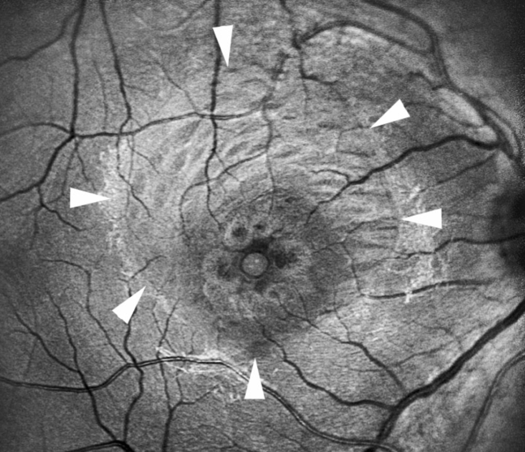
Dissociated optic nerve fiber layer appearance, seen as arcuate striae in the nerve fiber layer, is frequent after peeling and is usually of little visual consequence.
Small paracentral scotomas have been reported, and some studies show thinning of the inner retina and a change in the foveal contour.
Swelling of the arcuate nerve fiber layer and retinal hemorrhages can occur during the procedure, and most resolve.
The long-term importance of these changes remains an area of study.
Evidence and Controversies
A randomized trial in the year 2000 found a higher rate of macular hole closure when the membrane was peeled, and subsequent reviews reached similar conclusions for larger holes.
For small holes, studies suggest that closure rates are good even without peeling, and some surgeons choose not to peel to avoid trauma.
For epiretinal membranes, peeling may reduce recurrence but is not clearly beneficial in all eyes, and the risk of damage must be weighed.
Patients should be told that the surgery aims to close the hole or remove the membrane, that the visual result varies, and that distortion may persist for months.
Practical Tips for Surgeons and Trainees
Stain only the area that needs to be peeled and wash the dye out promptly to limit contact time.
Start the peel away from the fovea, and keep the flap close to the surface to avoid lifting the nerve fiber layer.
Avoid peeling beyond the vascular arcades, and keep the peeled area symmetric.
Many surgeons switch to a gas tamponade and positioning after peeling, and the choice depends on the hole size and the case.
Follow-Up After Peeling
Examine the macula with OCT at the first postoperative visit and at intervals until the hole is closed and the inner layers have settled.
Subtle changes, such as dimpling of the inner retina, are common and often resolve or remain asymptomatic.
Patients should be told that distortion and paracentral blur may improve slowly over many months.
Alternatives to full peeling include inverted flap techniques for large holes, in which a rim of membrane is left attached and folded over the hole as a scaffold.
These techniques have given good closure rates in large and myopic holes in several series, and they are increasingly used.


Document what you see
Two smartphone imaging tools built for everyday clinic use — one for the slit lamp, one for the fundus.
From Choroida — the team behind this siteReferences
- Brooks HL Jr. Macular hole surgery with and without internal limiting membrane peeling. Ophthalmology. 2000;107:1939-1948.
- Spiteri Cornish K, Lois N, Scott NW, et al. Vitrectomy with internal limiting membrane peeling versus no peeling for idiopathic full-thickness macular hole. Ophthalmology. 2014;121:649-655.
- Ryan SJ, Schachat AP, Wilkinson CP, et al, eds. Retina. 6th ed. Elsevier; 2018.
- American Academy of Ophthalmology. Basic and Clinical Science Course, Section 12: Retina and Vitreous. AAO; current edition.

Writes and reviews the clinical articles on Educate. Every post is checked against peer-reviewed sources before it goes live.