
Iridocorneal endothelial syndrome, abbreviated ICE syndrome, is best understood not as a single disease but as an umbrella term for three related presentations, Chandler syndrome, Cogan-Reese syndrome, and essential iris atrophy, that all share the same fundamental underlying abnormality: an abnormal population of corneal endothelial cells that migrates across the anterior chamber angle and iris surface, producing a spectrum of downstream effects that differ mainly in which structure bears the brunt of the process.
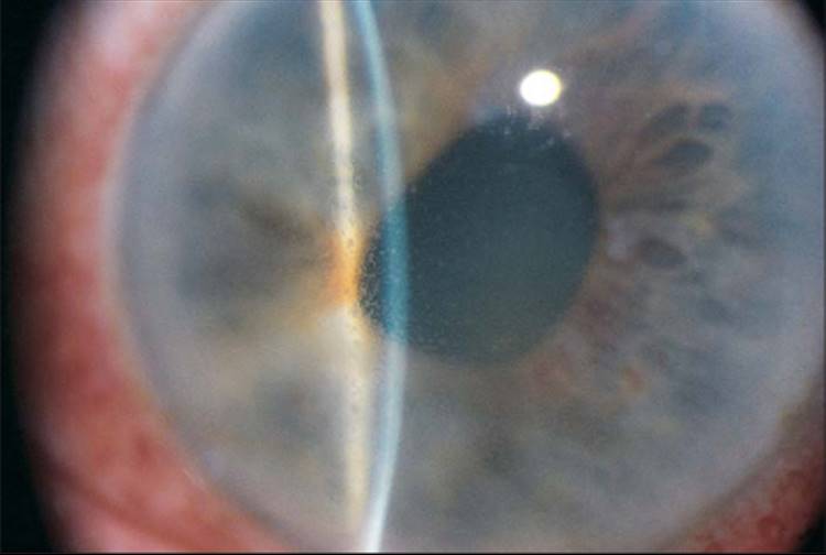
One Underlying Process, Three Clinical Faces
In all three ICE syndrome variants, an abnormal corneal endothelial cell population, behaving more like epithelial cells than typical endothelium, proliferates and spreads across the trabecular meshwork and iris surface, and it is the differing downstream consequences of this same abnormal cell migration and membrane formation, corneal changes, angle closure, and iris changes in varying proportions, that produce the three recognized clinical variants.
The Three Variants
Chandler Syndrome
Corneal endothelial and stromal changes, including corneal edema, predominate, with relatively mild iris changes, this being the most common of the three ICE syndrome variants (see Chandler syndrome for a detailed discussion of this specific variant).
Cogan-Reese Syndrome
Iris nodules, pigmented, pedunculated lesions on the iris surface, are the most distinctive feature, alongside variable degrees of corneal and angle involvement (see Cogan-Reese syndrome for a detailed discussion of this specific variant).
Essential Iris Atrophy
Progressive iris stromal thinning, corectopia (displacement of the pupil), and polycoria (the appearance of multiple pupils, from progressive iris hole formation) are the dominant features in this variant, with iris changes considerably more severe and disfiguring than typically seen in Chandler syndrome, though corneal and angle involvement still occur to varying degrees as part of the same underlying process.
All-fit Slit-Lamp Adapter
Record and share exactly what you see at the slit lamp. One adapter fits any slit lamp or surgical microscope — and any smartphone.
From Choroida — the team behind this siteShared Features Across All Three Variants
- Unilateral involvement is typical across all three variants, an important distinguishing feature from many other iris and corneal conditions that more commonly affect both eyes
- Middle-aged women are disproportionately affected, a demographic pattern shared across the ICE syndrome spectrum
- Secondary angle closure glaucoma, related to the abnormal membrane spreading across and eventually contracting over the trabecular meshwork and angle structures, is a significant potential complication shared across all three variants and often the most vision-threatening aspect of the disease
- Corneal edema, when present, relates to endothelial dysfunction from the abnormal cell population itself, again a feature shared to varying degrees across the three presentations
Diagnostic Evaluation
- Slit-lamp examination characterizing the specific pattern of corneal, angle, and iris findings present, supporting classification into the specific variant a given patient’s presentation most closely matches
- Specular or confocal microscopy, when available, can demonstrate the characteristic abnormal endothelial cell appearance shared across all ICE syndrome variants
- Gonioscopy to assess the degree of angle involvement and any associated peripheral anterior synechiae, relevant to both diagnosis and glaucoma risk assessment
- Intraocular pressure monitoring, given the shared risk of secondary angle closure glaucoma across all three variants
Management Principles Common to ICE Syndrome
Glaucoma Management
Secondary angle closure glaucoma, when present, is managed with a combination of medical therapy and, for more advanced or medically uncontrolled disease, surgical intervention, though surgical outcomes in ICE syndrome glaucoma can be more challenging than in primary glaucomas given the ongoing abnormal membrane activity that can affect surgical drainage pathways over time.
Managing Corneal Edema
When corneal edema develops from endothelial dysfunction, management follows similar principles to corneal endothelial decompensation from other causes, including consideration of endothelial keratoplasty for visually significant cases, though the ongoing, potentially progressive nature of the underlying abnormal endothelial process in ICE syndrome is a relevant consideration in long-term surgical planning and prognosis discussion.
Monitoring for Progression
Given that all three ICE syndrome variants can show gradual progression over time, periodic monitoring of corneal, angle, and iris findings, along with intraocular pressure, supports timely recognition and management of any progressive changes.
Practical points
Examine both eyes, since the condition is almost always unilateral and the fellow eye can serve as a comparison. Specular microscopy of the affected eye is a useful diagnostic test when the cornea is clear enough, and gonioscopy documents the synechiae.
Prognosis
Prognosis in ICE syndrome varies depending on the specific variant and degree of angle and corneal involvement, with the shared risk of secondary glaucoma representing the most significant threat to long-term visual function across all three presentations.
Understanding these three variants as related expressions of one underlying abnormal endothelial process, rather than as entirely separate diseases, supports a more coherent approach to monitoring and managing whichever specific pattern a given patient’s ICE syndrome presentation follows.


Document what you see
Two smartphone imaging tools built for everyday clinic use — one for the slit lamp, one for the fundus.
From Choroida — the team behind this siteReferences
- Shields MB. Progressive essential iris atrophy, Chandler’s syndrome, and the iris nevus (Cogan-Reese) syndrome: a spectrum of disease. Surv Ophthalmol. 1979;24:3-20.
- Campbell DG, Shields MB, Smith TR. The corneal endothelium and the spectrum of essential iris atrophy. Am J Ophthalmol. 1978;86:317-324.
- Laganowski HC, Kerr Muir MG, Hitchings RA. Glaucoma and the iridocorneal endothelial syndrome. Arch Ophthalmol. 1992;110:346-350.
- Alvarado JA, Underwood JL, Green WR, et al. Detection of herpes simplex viral DNA in the iridocorneal endothelial syndrome. Arch Ophthalmol. 1994;112:1601-1609.
