
Diabetic retinopathy in pregnancy needs a plan before conception, because pregnancy can accelerate existing retinopathy and treatment options are narrower once the patient is pregnant.
Most women with type 1 or type 2 diabetes who have little or no retinopathy at conception do well, whereas those with moderate to severe nonproliferative disease are the ones at real risk of progression.
The retina specialist’s job is to identify that risk early, coordinate with the obstetrician and diabetologist, and treat proliferative disease before it threatens vision.
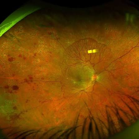
Why Pregnancy Affects the Retina
Several changes act together during pregnancy.
Blood flow and blood volume rise, and retinal autoregulation may be overwhelmed in eyes with existing capillary damage.
Hormonal and growth-factor changes, including placental growth factor and VEGF, increase vascular permeability.
Blood pressure may rise, particularly with preeclampsia, and hypertension is an independent driver of retinopathy progression.
Finally, tight glycemic control started rapidly in early pregnancy can produce an early worsening of retinopathy, similar to the early worsening seen in the DCCT with intensive therapy.
Who Is at Risk of Progression?
Progression is not uniform.
The main risk factors are:
- Retinopathy severity at conception, which is the strongest predictor
- Longer diabetes duration
- Poor glycemic control at conception
- A large and rapid fall in HbA1c during early pregnancy
- Hypertension, preeclampsia, or renal disease
- Type 1 diabetes, in which most of the evidence has been collected
In the Diabetes in Early Pregnancy Study, women with worse baseline retinopathy and higher initial HbA1c were more likely to progress, and rapid glycemic improvement was associated with early worsening.
Women with no retinopathy at conception rarely develop sight-threatening disease, although some develop mild changes.
Gestational diabetes does not appear to increase the risk of retinopathy, so routine retinal screening for gestational diabetes alone is generally not recommended.
Fundus Explorer Pro
Photograph the retinal findings described here with the phone already in your pocket — 22 D optics and built-in illumination in one handheld unit.
From Choroida — the team behind this siteScreening for Diabetic Retinopathy in Pregnancy
Current diabetes guidelines recommend a comprehensive eye examination before pregnancy or in the first trimester in women with preexisting type 1 or type 2 diabetes.
Follow-up is then tailored to severity, commonly every trimester and for a year after delivery.
Practical points for the examiner:
- Dilated fundus examination is safe in pregnancy
- OCT is safe and useful for macular edema
- Fluorescein angiography is generally avoided unless the information will change management
- Pregnant patients with no or mild retinopathy can be reviewed less often than those with moderate or worse disease
Women with severe nonproliferative or proliferative disease need closer follow-up, sometimes monthly.
Treatment During Pregnancy
Panretinal Photocoagulation
Laser is safe in pregnancy and remains the mainstay for high-risk proliferative disease.
It should be done promptly, ideally before or early in pregnancy, and the treatment can be split over sessions to limit macular edema (see panretinal photocoagulation).
Anti-VEGF Injections
VEGF is important for placental and fetal development, and systemic absorption of intravitreal agents has been documented.
Safety data in pregnancy are limited, so anti-VEGF injections are generally avoided and reserved for cases where the maternal benefit clearly outweighs the uncertain fetal risk.
This is a point to discuss with the obstetrician and to document.
Corticosteroids and Vitrectomy
Intravitreal or periocular steroids are used occasionally for center-involving macular edema that threatens vision, with limited data.
Vitrectomy for vitreous hemorrhage or tractional detachment is performed when needed, in coordination with anesthesia and obstetric care.
Macular Edema
Diabetic macular edema in pregnancy often improves after delivery, so observation is reasonable when vision allows and the patient can be followed closely.
Systemic Measures
Blood pressure should be controlled with pregnancy-safe agents, since ACE inhibitors and ARBs are contraindicated in pregnancy.
Statins and fenofibrate are also generally stopped, so some systemic measures used for retinopathy are not available.
Glycemic control should improve gradually, because an abrupt fall in HbA1c carries its own risk of early worsening.
Coordinating With the Obstetric Team
The retina specialist should send a written summary after each examination, stating the retinopathy grade, whether treatment is needed, and when the next review is due.
Delivery mode is decided by obstetric indications.
Stable nonproliferative or treated proliferative retinopathy is not by itself a reason to avoid vaginal delivery.
Active untreated proliferative disease or a recent vitreous hemorrhage deserves a discussion, because straining during labor raises intraocular venous pressure and can provoke hemorrhage.
There is limited evidence on this point, so the decision should be individualized and documented.
A history of rapid HbA1c reduction, new hypertension, or proteinuria should prompt earlier retinal review, because these changes tend to precede progression.
After Delivery
Retinopathy often stabilizes or partly regresses after delivery, particularly in mild to moderate disease.
Severe proliferative disease may not regress, and examination is recommended in the year after delivery.
Breastfeeding is compatible with laser, and the decision about anti-VEGF use after delivery depends on the medication and the infant.
Counseling Before Conception
Preconception counseling is one of the most effective interventions.
It includes:
- Dilated examination and treatment of any high-risk retinopathy before conception
- Glycemic control brought to target gradually
- Blood pressure control, with medication changes made before pregnancy
- A shared plan for screening intervals and who to call with visual symptoms
Patients should know the symptoms that warrant urgent review: sudden floaters, a curtain over the vision, and sudden central blur.
Preeclampsia and Other Retinal Problems
Retinal changes during pregnancy are not always diabetic.
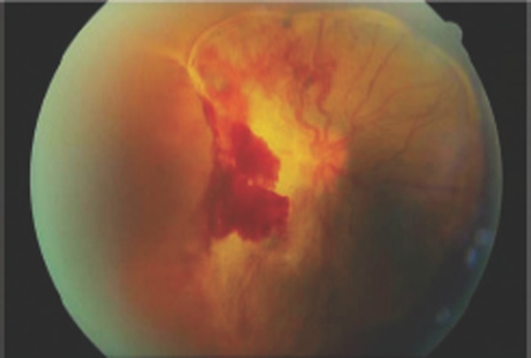
Preeclampsia and HELLP syndrome can cause arteriolar narrowing, cotton wool spots, serous retinal detachment, and cortical visual loss (see hypertensive retinopathy).
Central serous chorioretinopathy is also more common in the third trimester.
These conditions usually resolve after delivery, and they are separate from diabetic retinopathy even when they coexist.


Document what you see
Two smartphone imaging tools built for everyday clinic use — one for the slit lamp, one for the fundus.
From Choroida — the team behind this siteReferences
- Klein BE, Moss SE, Klein R. Effect of pregnancy on progression of diabetic retinopathy. Diabetes Care. 1990;13:34-40.
- Chew EY, Mills JL, Metzger BE, et al. Metabolic control and progression of retinopathy: the Diabetes in Early Pregnancy Study. Diabetes Care. 1995;18:631-637.
- The Diabetes Control and Complications Trial Research Group. Effect of pregnancy on microvascular complications in the Diabetes Control and Complications Trial. Diabetes Care. 2000;23:1084-1091.
- Rahman W, Rahman FZ, Yassin S, Al-Suleiman SA, Rahman J. Progression of retinopathy during pregnancy in type 1 diabetes mellitus. Clin Exp Ophthalmol. 2007;35:231-235.
- American Diabetes Association Professional Practice Committee. Standards of Care in Diabetes-2024. Diabetes Care. 2024;47(Suppl 1).
Test yourself
A few questions straight from this article.
-
Which factor most strongly predicts progression of diabetic retinopathy during pregnancy?
Severity at conception is the strongest predictor: women with little or no retinopathy generally do well, while moderate to severe non-proliferative disease carries real risk of progression. -
What retinal screening is recommended for gestational diabetes alone?
Gestational diabetes does not appear to raise the risk of retinopathy, so routine retinal screening for gestational diabetes alone is generally not recommended. -
Which change in early pregnancy is linked to early worsening of diabetic retinopathy?
Rapidly instituted tight glycaemic control can cause early worsening, mirroring the early worsening seen with intensive therapy in the DCCT, so control should improve gradually. -
When should a woman with pre-existing type 1 or type 2 diabetes have her eye examination for pregnancy?
Guidelines advise a comprehensive eye examination before pregnancy or in the first trimester, with follow-up then tailored to severity, commonly each trimester and for a year after delivery. -
Which investigation is generally avoided in a pregnant woman with diabetic retinopathy?
Dilated examination and OCT are both safe in pregnancy, whereas angiography is generally avoided unless the information will genuinely change management. -
What remains the mainstay of treatment for high-risk proliferative diabetic retinopathy in pregnancy?
Laser is safe in pregnancy and remains the mainstay for high-risk proliferative disease, done promptly and split over sessions to limit macular oedema. -
Why are anti-VEGF injections generally avoided during pregnancy?
VEGF matters for placental and fetal development and systemic absorption of intravitreal agents is documented, so with limited safety data these drugs are reserved for clear maternal benefit. -
How is centre-involving diabetic macular oedema often managed during pregnancy?
Macular oedema in pregnancy frequently improves once the pregnancy ends, so observation is reasonable when vision allows and close follow-up is possible. -
Which retinal findings point to preeclampsia or HELLP syndrome rather than diabetic retinopathy?
Preeclampsia and HELLP can produce arteriolar narrowing, cotton wool spots, serous retinal detachment and cortical visual loss, and these usually resolve after delivery. -
What typically happens to diabetic retinopathy after delivery?
Mild to moderate retinopathy often stabilises or partly regresses after delivery, while severe proliferative disease may not, so examination is advised during the following year.

Writes and reviews the clinical articles on Educate. Every post is checked against peer-reviewed sources before it goes live.


