
Primary corneal amyloidosis occupies a specific, important place in the differential diagnosis of corneal clouding: amyloid deposition confined to the cornea, without an identifiable systemic amyloid disease and without fitting the pattern of a defined hereditary corneal dystrophy like lattice dystrophy, and correctly placing a given patient’s findings into this category, rather than one of the other amyloid-related possibilities, shapes both the workup pursued and the counseling offered.
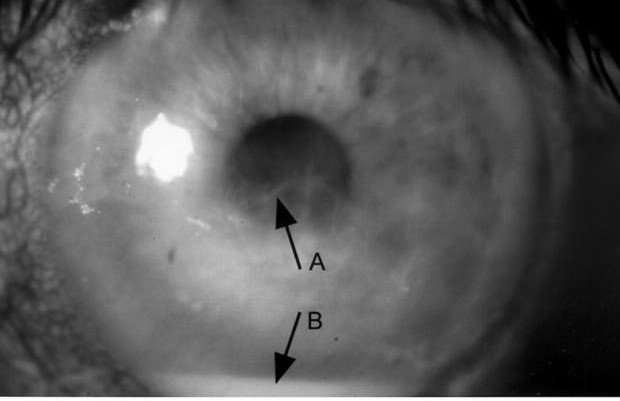
Amyloid in the Cornea: Several Different Stories
Amyloid deposition in the cornea is not a single diagnosis but rather a shared histologic finding that can arise through several distinct mechanisms, and distinguishing between them is the central diagnostic task whenever corneal amyloid is identified or suspected.
- Lattice corneal dystrophy, a specific, well-characterized hereditary condition producing the classic branching refractile lines, resulting from TGFBI gene mutations (see lattice corneal dystrophy)
- Gelatinous drop-like corneal dystrophy, a distinct hereditary condition producing dramatic, elevated, mulberry-shaped masses from a different genetic cause (see gelatinous drop-like corneal dystrophy)
- Secondary corneal amyloidosis, developing within a cornea chronically affected by another condition, including longstanding trachoma, chronic keratoconjunctivitis, or other prolonged ocular surface inflammatory or degenerative disease, where amyloid accumulates as a late, localized consequence of the underlying chronic process
- Primary (localized) corneal amyloidosis, in which amyloid deposition occurs within the cornea without an identifiable hereditary corneal dystrophy syndrome and without evidence of systemic amyloid disease, representing a more idiopathic, localized process
Clinical Presentation
- Gradual, progressive corneal clouding or the development of visible deposits, without the specific, recognizable pattern of lattice lines or gelatinous mulberry masses that would point to one of the named hereditary dystrophies
- Reduced vision correlating with the extent and density of the amyloid deposition
- Typically lacks the early, severe recurrent erosion prominent in some of the hereditary Bowman-layer and stromal dystrophies, though surface irregularity and mild erosion symptoms can still occur
- A relevant history of chronic ocular surface disease or prior significant ocular inflammation supports a secondary rather than primary process
All-fit Slit-Lamp Adapter
Record and share exactly what you see at the slit lamp. One adapter fits any slit lamp or surgical microscope — and any smartphone.
From Choroida — the team behind this siteDiagnostic Evaluation
- Careful slit-lamp examination to assess whether the deposition pattern matches a recognized hereditary dystrophy syndrome, since a pattern-matched diagnosis (lattice or gelatinous drop-like dystrophy) is generally more specific and informative than a default label of primary amyloidosis
- A thorough history for any chronic ocular surface disease, prior trachoma exposure, or longstanding keratoconjunctivitis that could explain a secondary process
- A family history, since hereditary corneal dystrophies are typically inherited in recognizable patterns, while primary localized amyloidosis without an underlying dystrophy syndrome is generally sporadic
- Biopsy or examination of surgically excised tissue with Congo red staining confirms the presence of amyloid and supports histologic characterization, though it does not on its own distinguish between the various possible underlying causes without correlation with the clinical picture and genetic testing when appropriate
- Systemic evaluation for amyloidosis is generally reserved for cases with other suggestive systemic findings, since isolated corneal amyloid deposition without a hereditary dystrophy pattern is usually a localized phenomenon rather than a marker of systemic disease
Management
Addressing Any Underlying Cause
When corneal amyloidosis is secondary to chronic ocular surface disease, managing that underlying condition is an important part of care, both for its own sake and to reduce ongoing stimulus for further amyloid accumulation.
Surgical Management of Visually Significant Deposits
- Phototherapeutic keratectomy can address more superficial amyloid deposits, improving corneal surface regularity and clarity
- Corneal transplantation, lamellar or penetrating depending on the depth and extent of involvement, is used for deeper, more visually significant deposition not adequately addressed by phototherapeutic keratectomy
Monitoring for Recurrence
As with the hereditary amyloid-related corneal dystrophies, localized recurrence of amyloid deposition after surgical treatment is possible, particularly when an underlying chronic secondary cause remains active, supporting ongoing follow-up after any surgical intervention.
Confirming the diagnosis
Amyloid shows apple-green birefringence under polarized light after Congo red staining, which helps in the pathologic diagnosis of removed tissue. Genetic testing is available for the common TGFBI-related and gelsolin-related forms. In a patient with lattice lines, ask about systemic features, particularly facial palsy and skin laxity in gelsolin amyloidosis, because these influence referral.
Prognosis
Prognosis depends substantially on correctly identifying which category of corneal amyloid deposition a given patient has, since secondary amyloidosis tied to an actively ongoing chronic surface disease may continue to progress or recur unless that underlying driver is controlled, while isolated, treated deposits without an ongoing stimulus often have a more stable long-term course.
Careful diagnostic categorization at the outset, distinguishing primary localized disease from a hereditary dystrophy syndrome or a secondary process with its own specific driver, supports the most accurate counseling and the most appropriately targeted management plan for each individual patient.


Document what you see
Two smartphone imaging tools built for everyday clinic use — one for the slit lamp, one for the fundus.
From Choroida — the team behind this siteReferences
- Rodrigues MM, Gaster RN, Pratt MV. Unusual immunoglobulin deposits in lattice corneal dystrophy. Invest Ophthalmol Vis Sci. 1981;20:124-132.
- Meisler DM, Fine M. Recurrence of the clinical signs of lattice corneal dystrophy (type I) in corneal transplants. Am J Ophthalmol. 1984;97:210-214.
- Klintworth GK. Corneal dystrophies. Orphanet J Rare Dis. 2009;4:7.
- Rummelt V, Meyer HJ, Naumann GO. Secondary corneal amyloidosis in interstitial keratitis of congenital lues. Br J Ophthalmol. 1992;76:246-248.
Test yourself
A few questions straight from this article.
-
Primary corneal amyloidosis is defined by amyloid deposition with which feature?
The term is reserved for amyloid confined to the cornea with no identifiable systemic amyloid disease and no defined hereditary corneal dystrophy pattern, making it a more idiopathic, localized process. -
Which gene underlies lattice corneal dystrophy, the hereditary cause of branching refractile corneal amyloid lines?
Lattice corneal dystrophy results from TGFBI gene mutations and produces the classic branching refractile lines, a pattern-matched diagnosis distinct from primary localized amyloidosis. -
Which corneal appearance characterises gelatinous drop-like corneal dystrophy?
Gelatinous drop-like dystrophy is a separate hereditary condition from a different genetic cause, producing dramatic elevated mulberry-shaped masses rather than lattice lines. -
Which history most supports secondary rather than primary corneal amyloidosis?
Secondary corneal amyloidosis develops in a cornea chronically affected by another disease, such as longstanding trachoma or chronic keratoconjunctivitis, with amyloid accumulating as a late local consequence. -
Which histological finding confirms amyloid in excised corneal tissue?
Congo red staining with apple-green birefringence under polarised light confirms amyloid, though it does not by itself distinguish which underlying cause produced the deposition. -
What inheritance pattern is expected in primary localized corneal amyloidosis?
Hereditary corneal dystrophies are inherited in recognisable patterns, whereas primary localized amyloidosis without an underlying dystrophy syndrome is generally sporadic, so family history helps separate them. -
When is systemic amyloidosis evaluation indicated in a patient with isolated corneal amyloid?
Isolated corneal amyloid without a hereditary dystrophy pattern is usually a localized phenomenon, so systemic work-up is reserved for patients with other suggestive systemic findings. -
Which procedure is used for more superficial, visually significant corneal amyloid deposits?
Phototherapeutic keratectomy addresses superficial amyloid and improves surface regularity and clarity; deeper, more extensive deposition requires lamellar or penetrating transplantation instead. -
After surgery for corneal amyloid deposits, what most favours local recurrence of deposition?
Localized recurrence after surgical treatment is possible and is particularly likely when an underlying chronic secondary cause remains active, which is why follow-up continues after any intervention. -
What most determines long-term prognosis in a patient with corneal amyloid deposition?
Prognosis depends substantially on correct categorisation: secondary disease with an active chronic driver may keep progressing or recurring, while isolated treated deposits without ongoing stimulus are often stable.
