
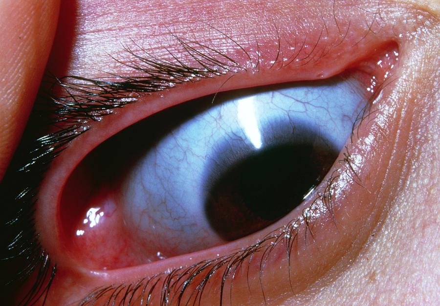
Osteogenesis imperfecta (OI) is a group of inherited disorders of type I collagen, characterized by bone fragility, and its most visible eye sign is a blue or gray tint of the sclera.
The eye is a collagen-rich organ, and the changes in the sclera and cornea can affect measurement of intraocular pressure and surgical safety.
Ophthalmologists see these patients for refraction, pressure checks, and surgery, so familiarity with the signs is worthwhile.
What Is Osteogenesis Imperfecta?
Most cases arise from mutations in the genes for the alpha chains of type I collagen, which cause a reduced or abnormal collagen.
The Sillence classification divides the disease into four types by severity and clinical features, and more types have been added as new genes were found.
Type I is the mildest and typically has blue sclera, while type II is lethal in the perinatal period, type III is progressively deforming, and type IV is moderate.
Other features include fractures with minimal trauma, short stature, dentinogenesis imperfecta, and hearing loss in adults.
Ocular Findings
- Blue sclera: thin, translucent sclera that reveals the underlying uvea, most obvious in type I
- Thin corneas: reduced central corneal thickness, associated with the presence of blue sclera
- Low ocular rigidity: a softer eye wall, which affects tonometry
- Keratoconus and megalocornea: reported with greater frequency in some series
- Glaucoma, cataract, and retinal detachment: described, but not common
- Embryotoxon and other anterior segment findings: occasional
The sclera is not always blue in other types, and the tint may fade with age in some patients.
All-fit Slit-Lamp Adapter
Record and share exactly what you see at the slit lamp. One adapter fits any slit lamp or surgical microscope — and any smartphone.
From Choroida — the team behind this siteEffect on Measurement of Pressure
Applanation tonometry underestimates the pressure in eyes with thin corneas.
A low ocular rigidity affects indentation tonometry, and Schiotz readings are unreliable.
Measure the central corneal thickness (see corneal pachymetry) and interpret Goldmann applanation readings accordingly (see Goldmann applanation tonometry).
Optic disc and visual field assessment should not be omitted, because a normal pressure reading may be falsely reassuring.
Differential Diagnosis of Blue Sclera
- Ehlers-Danlos syndrome: especially the kyphoscoliotic type (see Ehlers-Danlos syndrome)
- Marfan syndrome and Stickler syndrome: thin sclera in some patients
- Normal infants: a blue tint in the first year of life, when the sclera is thin
- Pseudoxanthoma elasticum and other connective tissue disorders: with different systemic findings
- Scleral thinning from ocular disease: scleritis, high myopia, or staphyloma
The combination of blue sclera, fractures, and dentinogenesis points toward OI.
Surgical Considerations
The sclera is fragile, and the risk of globe perforation during local anesthesia, strabismus surgery, and sutured procedures is higher.
Choose blunt needles and topical anesthesia when possible, and use care in suturing.
Cataract surgery is feasible, but corneal thinning and zonular weakness can complicate it.
Patients with OI can have difficult airways, anesthesia risks, and fracture risk during positioning, so coordination with the anesthesiologist is needed.
Follow-Up
Examine children for refractive error and amblyopia, and examine adults regularly for pressure, the optic disc, and the retina.
Patients should be informed that they need to protect their eyes from trauma and to report sudden changes in vision.
Care is often shared with a team that includes a pediatrician, an orthopedic surgeon, and a geneticist.
What Patients and Parents Ask
Parents often ask whether the blue color of the eyes is dangerous, and whether it suggests that the eyes are weak.
Explain that the color is due to the thinness of the outer wall of the eye, that most patients see normally, and that there is no treatment needed for the color.
Reassure them that vision is rarely affected by the blue sclera itself, and that the ophthalmologist’s role is to monitor refraction, pressure, and the health of the eye.
Pressure Measurement in Practice
When a patient with OI has a normal-looking disc and an intraocular pressure that seems normal, remember that the true pressure may be higher, and that the corneal correction factors are only approximate.
Use multiple measurements and, when possible, a dynamic contour or rebound tonometer, and follow the optic nerve and field closely.
A patient with a family history of glaucoma and OI deserves a more careful evaluation than the numbers alone suggest.
Hearing and Systemic Care
Hearing loss is common in adults with OI, and it may add to the difficulties of a patient with poor vision.
A multidisciplinary clinic helps to coordinate bisphosphonate therapy, orthopedic care, dental care, and hearing assessments.
Eye care professionals can contribute by communicating findings such as thin corneas to the team, and by asking about fractures in children with unexplained blue sclera.


Document what you see
Two smartphone imaging tools built for everyday clinic use — one for the slit lamp, one for the fundus.
From Choroida — the team behind this siteReferences
- Sillence DO, Senn A, Danks DM. Genetic heterogeneity in osteogenesis imperfecta. J Med Genet. 1979;16:101-116.
- Evereklioglu C, Madenci E, Bayazit YA, et al. Central corneal thickness is lower in osteogenesis imperfecta and negatively correlates with the presence of blue sclera. Ophthalmic Physiol Opt. 2002;22:511-515.
- Salmon JF. Kanski’s Clinical Ophthalmology: A Systematic Approach. 9th ed. Elsevier; 2020.
- American Academy of Ophthalmology. Basic and Clinical Science Course, Section 6: Pediatric Ophthalmology and Strabismus. AAO; current edition.

Writes and reviews the clinical articles on Educate. Every post is checked against peer-reviewed sources before it goes live.


