
Corneal pachymetry measures corneal thickness, a single number that quietly influences decisions across several unrelated areas of ophthalmology: how an intraocular pressure reading should be interpreted, whether a cornea is safe for refractive surgery, and how advanced a case of keratoconus or corneal oedema has become.
It is a simple measurement to obtain and easy to underuse, since its relevance is not always obvious at the point a patient is sitting in the chair for what looks like an unrelated reason.
Measurement Techniques
Ultrasound pachymetry remains the most widely used and validated method, using a handheld probe brought into direct contact with the anaesthetised corneal surface to measure thickness from the time delay of a reflected ultrasound pulse.
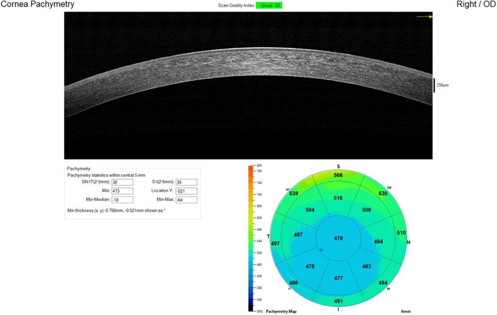
Optical pachymetry, including Scheimpflug imaging and anterior segment OCT, measures thickness non-contact, avoiding the need for topical anaesthesia and probe contact, and increasingly provides a full corneal thickness map rather than a single-point measurement.
Specular microscopy can also estimate corneal thickness as part of its broader assessment of the endothelium, though it is not typically used as the primary method when pachymetry alone is the clinical question.
Clinical Applications
Correcting Intraocular Pressure Interpretation
Goldmann applanation tonometry is calibrated for an assumed average corneal thickness, and eyes with thicker corneas tend to give falsely elevated pressure readings, while thinner corneas give falsely low ones relative to true intraocular pressure.
Central corneal thickness was established as an independent risk factor for progression from ocular hypertension to glaucoma in the Ocular Hypertension Treatment Study, making pachymetry a standard part of risk stratification for glaucoma suspects, not merely a correction factor applied to the pressure reading itself.
Refractive Surgery Planning
Adequate corneal thickness is required to safely perform LASIK or PRK while preserving a sufficient residual stromal bed, and pachymetry, combined with topography, is central to excluding patients at higher risk of post-refractive ectasia.
Keratoconus Monitoring
Progressive thinning is one of the parameters used to define disease progression in keratoconus, alongside changes in keratometry and refraction, and is directly relevant to the decision to proceed with corneal cross-linking.
Corneal Oedema and Endothelial Disease
Increased corneal thickness reflects endothelial dysfunction, as in Fuchs endothelial dystrophy or after intraocular surgery, and serial pachymetry is used to track the severity and course of corneal decompensation over time.
All-fit Slit-Lamp Adapter
Record and share exactly what you see at the slit lamp. One adapter fits any slit lamp or surgical microscope — and any smartphone.
From Choroida — the team behind this siteNormal Values and Interpretation
Average central corneal thickness in the general population is typically in the range of about 540 to 560 microns, though there is meaningful variation between individuals and some variation reported between different ethnic populations.
A minimum residual stromal bed of at least 250 microns is generally required after LASIK flap creation and ablation to maintain corneal biomechanical stability, which is the safety threshold pachymetry measurements are checked against during refractive surgery planning.
Progressive thinning on serial measurement, rather than a single absolute value, is often the more clinically meaningful finding in a condition like keratoconus, since it demonstrates an active, ongoing process rather than a static baseline characteristic of that individual cornea.
Practical Considerations
Measurements should be taken at a consistent time of day when tracking a patient serially, since corneal thickness shows a mild diurnal variation, typically slightly thicker on waking and thinning through the day as the eyes stay open.
Multiple readings are often averaged to improve reliability, particularly with ultrasound pachymetry, where probe angle and exact point of contact can introduce some measurement variability between individual readings.
Comparing measurements from different devices or different techniques over time should be done cautiously, since ultrasound and optical methods do not always agree exactly, and switching technique partway through a patient’s follow-up can introduce an apparent change that is really just a difference between instruments rather than a true clinical change.
Clinical Impact
Pachymetry has become integrated into routine glaucoma risk assessment, refractive surgery screening protocols, and keratoconus monitoring, functioning less as a standalone diagnostic test and more as essential context that changes how other measurements and decisions in each of these areas are interpreted.
Its main clinical value lies less in the number itself and more in what that number changes about the interpretation of other findings, whether that is trusting an intraocular pressure reading, clearing a cornea for laser surgery, or confirming that a keratoconic cornea is genuinely progressing rather than simply being re-measured with some normal variability.


Document what you see
Two smartphone imaging tools built for everyday clinic use — one for the slit lamp, one for the fundus.
From Choroida — the team behind this siteReferences
- Gordon MO, Beiser JA, Brandt JD, et al. The Ocular Hypertension Treatment Study: baseline factors that predict the onset of primary open-angle glaucoma. Archives of Ophthalmology. 2002.
- Doughty MJ, Zaman ML. Human corneal thickness and its impact on intraocular pressure measures. Survey of Ophthalmology. 2000.
- Wolffsohn JS, Safeen S, Shah S, Laiquzzaman M. Changes of corneal biomechanics with keratoconus. Cornea. 2012.
- Corneal Pachymetry. EyeWiki, American Academy of Ophthalmology.
- Pachymetry. StatPearls, NCBI Bookshelf.
Test yourself
A few questions straight from this article.
-
How does ultrasound pachymetry determine corneal thickness?
A handheld probe placed on the anaesthetised cornea measures thickness from the time delay of the reflected pulse, and remains the most widely used and validated method. -
What advantage do optical pachymetry methods such as anterior segment OCT offer?
Scheimpflug imaging and anterior segment OCT measure without contact or topical anaesthesia, and increasingly give a full corneal thickness map rather than a single point. -
What did the Ocular Hypertension Treatment Study establish about central corneal thickness?
The study showed central corneal thickness independently predicts progression from ocular hypertension to glaucoma, so pachymetry is risk stratification, not merely a correction factor. -
What is the typical average central corneal thickness in the general population?
Average central corneal thickness sits around 540 to 560 microns, with meaningful variation between individuals and some reported variation between ethnic populations. -
What minimum residual stromal bed is generally required after LASIK flap creation and ablation?
A residual stromal bed of at least 250 microns is generally required to maintain corneal biomechanical stability, and is the threshold checked during refractive planning. -
Which pachymetry finding in keratoconus is directly relevant to the decision to proceed with cross-linking?
Progressive thinning, alongside changing keratometry and refraction, defines progression in keratoconus and bears directly on the decision to cross-link. -
Increased corneal thickness on pachymetry most directly reflects which underlying problem?
Thickening reflects endothelial dysfunction, as in Fuchs endothelial dystrophy or after intraocular surgery, and serial pachymetry tracks the course of decompensation. -
Why should serial pachymetry measurements be taken at a consistent time of day?
The cornea is typically slightly thicker on waking and thins through the day while the eyes stay open, so consistent timing matters when tracking a patient. -
Why are multiple readings usually averaged when performing ultrasound pachymetry?
Probe angle and the exact point of contact vary between readings, so averaging several measurements improves reliability with ultrasound pachymetry. -
Why should measurements from different pachymetry devices be compared cautiously over time?
Ultrasound and optical methods do not always agree exactly, so switching technique mid-follow-up can create an apparent change that is really an instrument difference.

Writes and reviews the clinical articles on Educate. Every post is checked against peer-reviewed sources before it goes live.


