
Glaukomflecken, or “glaucoma flecks,” are small, gray-white, patchy opacities in the anterior subcapsular zone of the lens, usually located in the pupillary area.
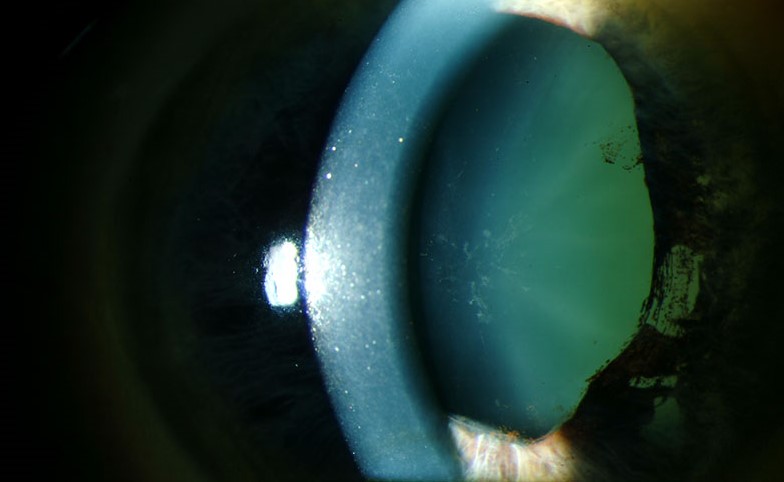
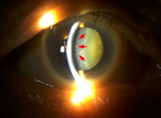
They are a marker of a previous episode of acute angle closure, and they are valuable because they show that the eye has had a severe, often unrecognized, rise in intraocular pressure.
How they form
During an acute attack, intraocular pressure rises abruptly to very high levels, often over 50 mmHg.
The lens epithelium, which lies directly beneath the anterior capsule, is injured by the ischemia and pressure, and by the damage to the aqueous that bathes it.
The injured cells die, and the underlying lens fibers become opaque.
Over weeks, new fibers are laid down beneath the damaged area, and the opacity is covered by clear lens substance and appears to be moving deeper into the lens.
Appearance
- Multiple small gray-white flecks, often in a patchy or wedge-shaped distribution
- Located just beneath the anterior capsule in the central or pupillary zone, since this is the area most exposed to aqueous and the zone that is not covered by the iris in a mid-dilated pupil
- Often found with other signs of past angle closure, including sector iris atrophy, a slightly dilated, irregular, poorly reactive pupil, and peripheral anterior synechiae on gonioscopy
- Persist for life and move deeper with time as new fibers develop
All-fit Slit-Lamp Adapter
Record and share exactly what you see at the slit lamp. One adapter fits any slit lamp or surgical microscope — and any smartphone.
From Choroida — the team behind this siteClinical significance
Glaukomflecken do not affect vision, and they do not need treatment.
They serve as a time-stamped record that an eye has had a significant angle closure attack.
That has implications for the fellow eye, which carries a high risk of an attack without treatment, and for the affected eye, which may need further treatment for chronic angle closure.
Differential diagnosis
Anterior subcapsular opacities occur in other conditions: atopic dermatitis (shield-like cataract), trauma, uveitis, and chronic exposure to certain drugs.
The combination of a history of eye pain, halos, and the other angle closure signs in the same eye makes the diagnosis clear.
What to do when you see them
- Examine the angle by gonioscopy in both eyes
- Measure intraocular pressure and assess the optic nerve and visual field
- Look for iris atrophy and posterior synechiae
- Treat accordingly: laser peripheral iridotomy for the fellow eye, and for the affected eye, treatment of any remaining angle closure or glaucoma (see Nd:YAG laser peripheral iridotomy)
- Consider early lens extraction when the angle remains narrow, and cataract is present
Patient counselling
Patients are often surprised to learn that they have already had an attack.
They should know the symptoms of an acute episode, which are severe pain, redness, blurred vision, and halos around lights, and they should be told to seek emergency care for these signs.
Early treatment saves vision.
Why the flecks are located where they are
After an acute attack, ischemia and the toxic effects of high pressure kill lens epithelial cells beneath the anterior capsule in the area exposed through the pupil, which is the central zone of the lens. The flecks therefore tend to lie in the center and in a ring that corresponds to the diameter of the mid-dilated pupil during the attack. Later, as the lens epithelium forms new fibers, the flecks are buried deeper, and they may become less distinct.
Other signs of an old attack
Look for sectoral iris atrophy, a pupil that is slightly dilated and irregular, a scarred or wavy iris margin, peripheral anterior synechiae on gonioscopy, and pigment on the corneal endothelium. A history of episodes of eye pain, headache, nausea, and halos may be present, and the patient may have attributed them to migraine. Ask about these symptoms specifically.
What it means for the fellow eye
The fellow eye shares the anatomical predisposition to angle closure and needs a prophylactic laser peripheral iridotomy in most cases. Explain to the patient that this is a quick outpatient procedure that greatly reduces the chance of an attack, and give advice about the warning symptoms. Patients who have had an attack should avoid medications that dilate the pupil, such as some antihistamines, anticholinergics, and decongestants, unless their eyes have been treated.
Documentation
When the flecks are found, record their location and size, the pupil appearance, the angle findings, and the intraocular pressure. These notes help to document the history of an attack that the patient may not remember, and they support the decision to treat the fellow eye.
Prognosis
The flecks are benign.
The prognosis of the eye depends on the extent of any optic nerve damage from the previous episode, and on control of the angle and pressure in the future.


Document what you see
Two smartphone imaging tools built for everyday clinic use — one for the slit lamp, one for the fundus.
From Choroida — the team behind this siteReferences
- Weinreb RN, Aung T, Medeiros FA. The pathophysiology and treatment of glaucoma: a review. JAMA. 2014;311:1901-1911.
- Sun X, Dai Y, Chen Y, et al. Primary angle closure glaucoma: what we know and what we don’t know. Prog Retin Eye Res. 2017;57:26-45.
- Saw SM, Gazzard G, Friedman DS. Interventions for angle-closure glaucoma: an evidence-based update. Ophthalmology. 2003;110:1869-1878.
- Duke-Elder S. System of Ophthalmology. Vol XI: Diseases of the Lens and Vitreous; Glaucoma and Hypotony. London: Kimpton; 1969.
