Optic nerve hypoplasia (ONH) is a congenital underdevelopment of the optic nerve, with fewer axons than normal, and it is one of the most common causes of poor vision in children in developed countries.
The visual outcome ranges from normal acuity to no light perception, and the condition may affect one or both eyes.
The eye finding matters beyond vision, since ONH is often associated with midline brain anomalies and pituitary hormone deficiencies that can be life-threatening if missed.
Every child with ONH needs a systemic evaluation that goes beyond the eye examination.
What Is Optic Nerve Hypoplasia?
ONH results from incomplete development of retinal ganglion cell axons during fetal life.
The optic disc is smaller than normal, and the nerve fiber layer is thin.
Several associations have been identified.
- Young maternal age and first pregnancy
- Maternal diabetes
- Maternal alcohol, drug, and medication exposure during pregnancy
- Genetic variants involving transcription factors such as HESX1, SOX2, and SOX3 in a minority
- Prematurity and low birth weight
The cause is often unknown, and most cases are sporadic.
When ONH occurs with absence of the septum pellucidum, corpus callosum abnormalities, and pituitary dysfunction, the combination is called septo-optic dysplasia (see septo-optic dysplasia).
Clinical Features
Visual Function
Visual acuity ranges widely, and it does not always match the appearance of the disc.
Bilateral cases commonly have poor vision and searching nystagmus, and parents may notice that the infant does not fix and follow.
Unilateral cases may present with strabismus or leukocoria-like appearance of the pupil, and some are found at a routine screening.
Visual field defects are common, and may be generalized constriction or arcuate defects.
The Disc
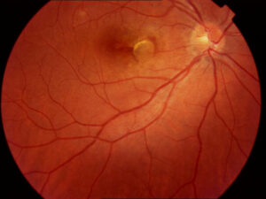
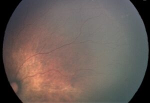
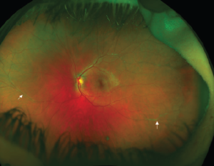
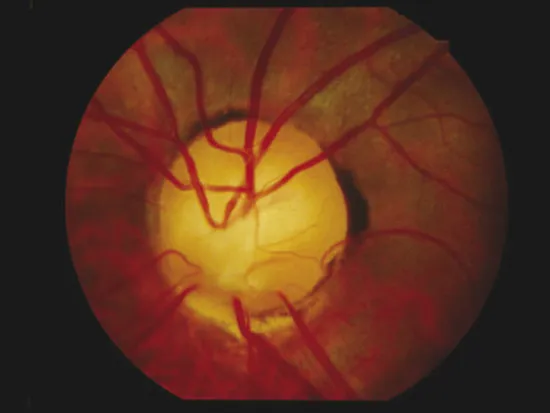
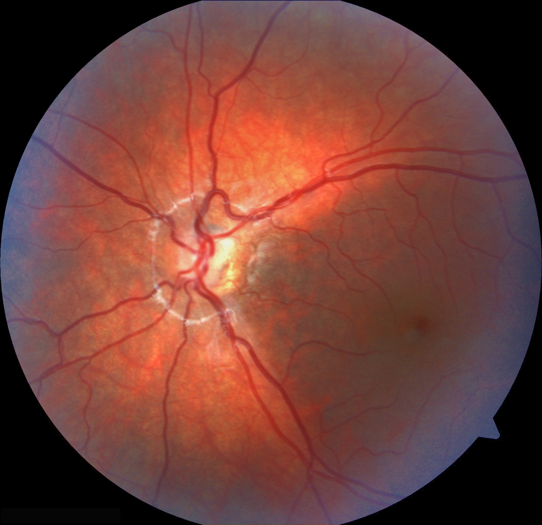
The optic disc is small, gray or pale, and often surrounded by a yellowish peripapillary halo bordered by a ring of pigment.
This produces the double ring sign, in which the outer ring marks the normal scleral canal and the inner ring marks the actual neural rim.
Retinal vessels are often tortuous, and the disc may look normal in mild cases.
The ratio between the distance from the disc to the fovea and the disc diameter exceeds about three in many eyes with ONH, which helps when the diagnosis is uncertain.
Other Ocular Findings
- Nystagmus in bilateral severe cases
- Strabismus and amblyopia
- Refractive error, often astigmatism or myopia
- Foveal hypoplasia in some patients (see foveal hypoplasia)
- Peripapillary staphyloma and microphthalmia in rare cases
Fundus Explorer Pro
Photograph the retinal findings described here with the phone already in your pocket — 22 D optics and built-in illumination in one handheld unit.
From Choroida — the team behind this siteSystemic Associations
Pituitary Hormone Deficiency
Hypopituitarism is the most important systemic association.
- Growth hormone deficiency is the most frequent, and it may lead to short stature and neonatal hypoglycemia
- ACTH deficiency causes hypocortisolism, which is dangerous during illness and surgery
- Thyroid hormone deficiency affects growth and development
- Antidiuretic hormone deficiency (diabetes insipidus) may occur
- Sex hormone abnormalities and early or delayed puberty
Deficiencies may appear over time, so a normal endocrine profile in infancy does not exclude later development.
Brain Anomalies
MRI may show absence of the septum pellucidum, thin corpus callosum, hypoplastic optic nerves and chiasm, ectopic posterior pituitary, and cortical malformations such as schizencephaly.
Developmental delay, cerebral palsy, and epilepsy are common in some children.
Investigations
- Dilated fundus examination with photographs to document disc size and appearance
- OCT of the peripapillary RNFL and macula, which shows thin RNFL and helps to assess the fovea
- MRI of the brain and pituitary to define optic nerves, chiasm, pituitary, and midline structures
- Endocrine assessment with growth measurements, cortisol, thyroid function, IGF-1, prolactin, and referral to pediatric endocrinology
- Visual evoked potentials or ERG when visual function is difficult to assess
- Genetic testing in selected patients
A child with ONH and hypoglycemia, prolonged jaundice, or poor growth should be referred urgently.
Differential Diagnosis
- Optic atrophy of other causes (see optic atrophy)
- Morning glory disc anomaly and coloboma (see morning glory syndrome and coloboma)
- Tilted disc syndrome and myopic optic discs that look small (see tilted disc syndrome)
- Glaucomatous cupping
- Oligomeganephronia and other conditions with small optic nerves
- Delayed visual maturation
Careful measurement helps, since a small disc with normal function is a normal variant.
Management
There is no treatment that restores axons, so management centers on optimizing the vision that exists and on addressing systemic problems.
Visual Rehabilitation
- Correct refractive error with glasses early
- Treat amblyopia with patching or other measures in unilateral or asymmetric cases, since some improvement is possible (see amblyopia)
- Manage strabismus and nystagmus as indicated
- Refer for low-vision services and educational support
- Provide developmental and orientation-and-mobility services
Systemic Care
- Coordinate with pediatric endocrinology for regular screening and hormone replacement
- Inform anesthetists and emergency teams about the risk of adrenal insufficiency
- Involve neurology and developmental pediatrics when appropriate
Family Counseling
Parents need clear information that ONH is congenital, is not caused by anything they did, and does not progress.
Visual potential is difficult to predict in infancy, and acuity often improves as the child matures.
Prognosis
Vision is stable and does not worsen.
Many children with unilateral ONH have good function in the other eye and lead normal lives.
Children with bilateral severe ONH may have significant visual impairment and need long-term support.
The systemic prognosis depends on the presence of pituitary and brain abnormalities, and regular monitoring prevents complications such as hypoglycemia and adrenal crisis.


Document what you see
Two smartphone imaging tools built for everyday clinic use — one for the slit lamp, one for the fundus.
From Choroida — the team behind this siteReferences
- Borchert M. Reappraisal of the optic nerve hypoplasia syndrome. J Neuroophthalmol. 2012;32:58-67.
- Garcia-Filion P, Borchert M. Optic nerve hypoplasia syndrome: a review of the epidemiology and clinical associations. Curr Treat Options Neurol. 2013;15:78-89.
- Patel L, McNally RJ, Harrison E, Lloyd IC, Clayton PE. Geographical distribution of optic nerve hypoplasia and septo-optic dysplasia in Northwest England. J Pediatr. 2006;148:85-88.
- Ahmad T, Garcia-Filion P, Borchert M, et al. Endocrinological and auxological abnormalities in young children with optic nerve hypoplasia: a prospective study. J Pediatr. 2006;148:78-84.
- Brodsky MC. Pediatric Neuro-Ophthalmology. 3rd ed. New York: Springer; 2016.
Test yourself
A few questions straight from this article.
-
What underlies optic nerve hypoplasia?
ONH is a congenital underdevelopment of the optic nerve with fewer axons than normal, arising from incomplete development of retinal ganglion cell axons during fetal life, leaving a small disc and thin nerve fibre layer. -
What constitutes the double ring sign seen in optic nerve hypoplasia?
The small grey disc is surrounded by a yellowish peripapillary halo bordered by pigment: the outer ring marks the normal scleral canal while the inner ring marks the actual neural rim. -
Which measurement supports a diagnosis of optic nerve hypoplasia when the disc appearance is uncertain?
The ratio of the disc-to-fovea distance to the disc diameter exceeds about three in many eyes with ONH, which is useful when the diagnosis is uncertain. -
Which combination of findings with optic nerve hypoplasia is called septo-optic dysplasia?
When ONH occurs with absence of the septum pellucidum, corpus callosum abnormalities and pituitary dysfunction, the combination is termed septo-optic dysplasia. -
Which pituitary hormone deficiency is most frequent in children with optic nerve hypoplasia?
Growth hormone deficiency is the most frequent endocrine abnormality in ONH, and it may lead to short stature and neonatal hypoglycaemia. -
Why must anaesthetic and emergency teams be told that a child has optic nerve hypoplasia?
ACTH deficiency causes hypocortisolism, which is dangerous during illness and surgery, so anaesthetists and emergency teams should be informed of the risk of adrenal insufficiency. -
A child with optic nerve hypoplasia has normal hormone levels in infancy. What follows?
Pituitary deficiencies may appear over time, so a normal endocrine profile in infancy does not exclude later development and regular screening with paediatric endocrinology is needed. -
How is optic nerve hypoplasia distinguished from a physiologically small optic disc?
Careful measurement helps in the differential diagnosis, because a small disc with normal visual function is a normal variant rather than hypoplasia. -
Which MRI finding is characteristically sought in a child with optic nerve hypoplasia?
MRI of brain and pituitary may show an ectopic posterior pituitary, absence of the septum pellucidum, a thin corpus callosum, hypoplastic nerves and chiasm, and cortical malformations such as schizencephaly. -
What does management of optic nerve hypoplasia consist of?
No treatment restores axons, so care centres on refractive correction, amblyopia therapy, low-vision and educational support, and on endocrine and neurological care; vision itself is stable and does not worsen.

Writes and reviews the clinical articles on Educate. Every post is checked against peer-reviewed sources before it goes live.