
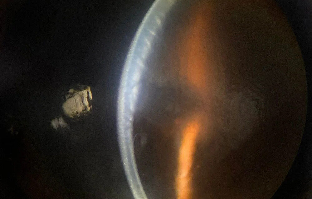
A cornea that has lost its clarity and become thick is one of the most common problems in anterior segment practice.
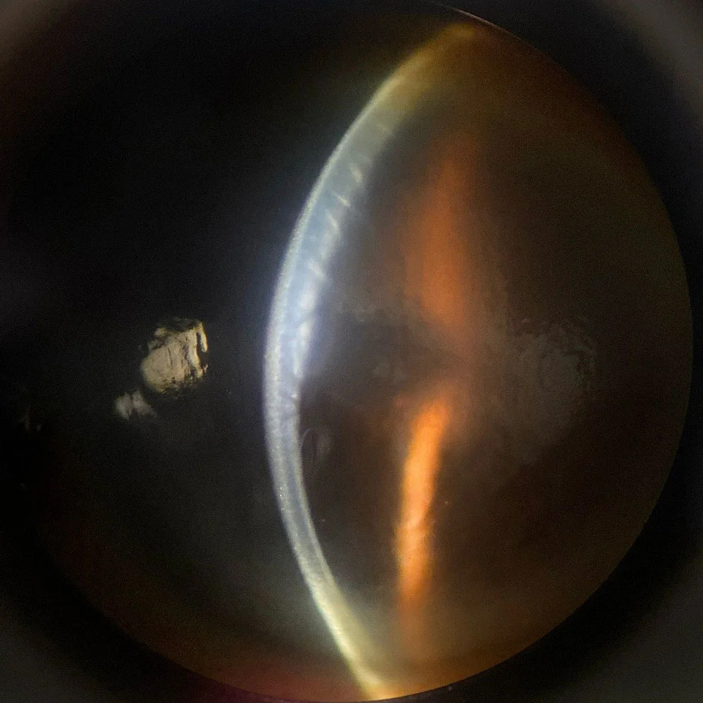
The reason is simple: the cornea stays clear because the endothelium pumps water out of the stroma.
If the pump fails, if the barrier is breached, or if the pressure in the eye is so high that fluid is driven in, the cornea swells.
The cause determines the treatment, so the first task is to sort it out.
Start with the layer
Endothelial failure
The commonest and most important cause.
Endothelial cells do not regenerate in humans, so a loss in cell number or function is permanent unless it is treated.
- Fuchs endothelial corneal dystrophy
- Pseudophakic and aphakic bullous keratopathy after intraocular surgery (see pseudophakic bullous keratopathy and aphakic bullous keratopathy)
- Iridocorneal endothelial syndrome and posterior polymorphous dystrophy (see ICE syndrome)
- Trauma, including birth trauma with Descemet tears
- Toxic anterior segment syndrome and chemical injury (see TASS)
- Uveitis and endotheliitis from herpes viruses or cytomegalovirus
Pressure-related
- Acute angle closure and other causes of very high intraocular pressure drive fluid into the epithelium, producing a steamy cornea with microcystic edema
- Congenital glaucoma, in which Haab striae may be present (see Haab striae)
Epithelial hypoxia
- Contact lens overwear, producing Sattler veil (see Sattler veil)
- Tight lenses, sleeping in lenses, or abnormal tear exchange
Stromal inflammation or structural causes
- Herpetic disciform keratitis and other keratitis (see herpetic stromal keratitis)
- Acute hydrops in keratoconus (see corneal hydrops)
- Descemet membrane detachment after surgery (see Descemet membrane detachment)
- Congenital hereditary endothelial dystrophy (see CHED)
Questions that narrow the list
- Is it unilateral or bilateral? Bilateral points to dystrophy or systemic factors, unilateral points to trauma, surgery, glaucoma, or inflammation.
- How quickly did it develop? Sudden onset favors acute glaucoma, hydrops, or trauma. Gradual onset with morning blur favors Fuchs.
- Is it painful and red? Pain suggests an acute process or epithelial breakdown.
- Is there a history of intraocular surgery, contact lens use, or eye inflammation?
- What is the intraocular pressure?
All-fit Slit-Lamp Adapter
Record and share exactly what you see at the slit lamp. One adapter fits any slit lamp or surgical microscope — and any smartphone.
From Choroida — the team behind this siteExamination
- Slit-lamp examination to see where the edema is: epithelial microcysts and bullae, stromal thickening and folds, or both
- Intraocular pressure measurement
- Examination of the anterior chamber for cells, flare, keratic precipitates, and the angle
- Pachymetry to quantify the thickness and follow it
- Specular microscopy or corneal tomography when the view allows, to examine the endothelium, with guttae in Fuchs dystrophy
Treatment
- Treat the cause first: lower the pressure in angle closure, stop the contact lens, control uveitis, or give antivirals as appropriate
- Hypertonic saline drops and ointment reduce epithelial edema
- Lowering the intraocular pressure reduces the force driving fluid in
- Bandage contact lens relieves pain in bullous keratopathy
- Endothelial keratoplasty (DMEK or DSAEK) replaces the failing endothelium, and is the usual surgical option today (see corneal endothelial decompensation)
- In select cases, Descemet stripping with ROCK inhibitor treatment is used
A practical way to localize the problem
Look at the cornea in three steps. First, check the epithelium for microcysts and bullae, which signal that the fluid has reached the surface. Second, check the stroma for thickening and folds. Third, look at the endothelium with specular reflection or a narrow beam for guttae, pigment, keratic precipitates, and Descemet folds. Compare the pachymetry values with the normal range, and with the other eye.
When edema is a surgical emergency
Acute angle closure, a Descemet detachment after surgery, and a corneal infection with melting need urgent attention. A cloudy cornea with a mid-dilated pupil and pain must be treated as angle closure until the pressure has been measured. A cloudy cornea after intraocular surgery should prompt an examination of the anterior chamber for an attached Descemet membrane.
What to tell the patient with chronic edema
Patients with endothelial failure often wake up with blurred vision that gradually improves during the day, as the cornea dehydrates by evaporation. Explain that this is a typical pattern, that drops and a hairdryer held at arm's length may provide temporary relief, and that the definitive treatment is a transplant of endothelium when the vision or comfort is affected. Describe the success rates and the follow-up involved so that the decision is informed.
Follow-up measures
Monitor corneal thickness and visual acuity over time. In endothelial disease, repeated measurements of central corneal thickness and a record of the time of day give an idea of progression. When surgery is under discussion, describe the options and expected recovery so that the patient can plan.
Prognosis
Edema that is due to a reversible cause often recovers fully.
Edema from permanent endothelial failure requires transplant for lasting clearing, and visual outcomes after modern endothelial keratoplasty are excellent in most patients.


Document what you see
Two smartphone imaging tools built for everyday clinic use — one for the slit lamp, one for the fundus.
From Choroida — the team behind this siteReferences
- Waring GO 3rd, Bourne WM, Edelhauser HF, Kenyon KR. The corneal endothelium. Normal and pathologic structure and function. Ophthalmology. 1982;89:531-590.
- Eghrari AO, Riazuddin SA, Gottsch JD. Fuchs corneal dystrophy. Prog Mol Biol Transl Sci. 2015;134:79-97.
- Krachmer JH, Mannis MJ, Holland EJ, eds. Cornea. 3rd ed. Philadelphia: Elsevier; 2011.
- Price MO, Mehta JS, Jurkunas UV, Price FW Jr. Corneal endothelial dysfunction: evolving understanding and treatment options. Prog Retin Eye Res. 2021;82:100904.
