
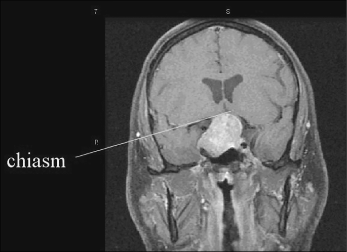
Chiasmal compression syndrome describes the characteristic pattern of visual loss that develops when a mass lesion — most commonly a pituitary adenoma — compresses the optic chiasm.
Its signature finding, bitemporal hemianopia, is one of the most anatomically satisfying localizations in all of neuro-ophthalmology, because it points almost unerringly to a single small region of the brain.
Recognizing this pattern early matters enormously, because many of these lesions are treatable and vision can be substantially recovered if compression is relieved before permanent optic nerve damage accumulates.
Why Bitemporal Hemianopia Happens
At the optic chiasm, nerve fibers from the nasal retina of each eye — which carry visual information from the temporal (outer) visual field — cross to the opposite side, while fibers from the temporal retina remain uncrossed.
A lesion compressing the chiasm from below or within, as a pituitary tumor typically does, preferentially affects these crossing nasal fibers, producing loss of the temporal visual field in both eyes while the nasal fields, served by the uncrossed fibers, are spared.
This crossing anatomy is the entire reason the visual field defect takes on such a specific, bilateral, symmetric pattern rather than a more generic, unilateral loss.
Common Causes
- Pituitary adenoma — the most common cause overall, whether hormonally functioning (with associated endocrine symptoms) or non-functioning (presenting with visual symptoms alone, sometimes as the first clue to the tumor’s presence)
- Craniopharyngioma — more common in children and young adults, often with additional hypothalamic and endocrine involvement given its typical suprasellar location
- Meningioma of the tuberculum sellae or diaphragma sellae
- Aneurysm of the anterior communicating or internal carotid artery, compressing the chiasm from an adjacent location
- Rarely, metastatic disease or other suprasellar tumors
Fundus Explorer Pro
Photograph the retinal findings described here with the phone already in your pocket — 22 D optics and built-in illumination in one handheld unit.
From Choroida — the team behind this siteClinical Presentation
Vision loss from chronic chiasmal compression is typically gradual and can be surprisingly well tolerated for a long time, because bitemporal field loss does not affect central acuity in early stages and patients often adapt without realizing how much peripheral field they have lost.
A classic and frequently cited example is patients only recognizing their visual field defect when it is specifically pointed out, sometimes after being involved in a car accident related to missed peripheral traffic.
Endocrine symptoms (menstrual irregularity, galactorrhea, symptoms of hormone excess or deficiency, or symptoms of hypopituitarism) may accompany the visual complaint and are worth asking about specifically, since a pituitary lesion is by far the most likely underlying cause.
Exam Findings
- Bitemporal hemianopia on formal visual field testing — classically starting superiorly (from inferior chiasmal compression, typical of a pituitary adenoma growing upward from below) and progressing to involve the full temporal field bilaterally as compression worsens
- Reduced visual acuity in more advanced or asymmetric compression
- Optic disc pallor, often with a distinctive “band” or “bowtie” pattern of atrophy corresponding to the specific nerve fiber bundles carrying the crossing fibers, visible on careful fundus exam in longstanding cases
- A relative afferent pupillary defect if compression is significantly asymmetric between the two optic nerves, discussed in more detail in this site’s dedicated article on that sign
- Occasionally, other findings from local mass effect — diplopia from cavernous sinus extension affecting the ocular motor nerves, or signs of raised intracranial pressure with larger lesions
Differential Diagnosis
- Bilateral optic neuropathy from other causes (toxic, nutritional, hereditary) — produces central or cecocentral field defects rather than the specific bitemporal pattern, discussed in this site’s separate coverage of toxic and nutritional optic neuropathy
- Retinitis pigmentosa or other causes of peripheral field constriction — a concentric rather than hemianopic pattern, and with characteristic fundus findings
- Homonymous hemianopia from a retrochiasmal lesion — affects the same side of the visual field in both eyes rather than the temporal field of each eye, localizing to the optic tract or brain rather than the chiasm itself
Diagnostic Evaluation
Formal automated visual field testing (Humphrey or similar) confirms and precisely characterizes the pattern of loss, and MRI of the brain and pituitary with attention to the sellar and suprasellar region identifies the underlying mass lesion.
Endocrine evaluation, including pituitary hormone panels, is essential once a sellar mass is identified, because both hormone-secreting tumors and hypopituitarism from a non-functioning mass require their own specific management alongside whatever is done for the visual findings.
Management
Surgical decompression — most often via a transsphenoidal approach for pituitary adenomas — is the primary treatment for vision-threatening chiasmal compression.
Visual recovery after decompression can be substantial, particularly when surgery occurs before longstanding, severe optic atrophy has developed, which is the central reason timely diagnosis matters so much in this condition, since the visual field defect’s duration and severity at the time of treatment are among the strongest predictors of how much vision ultimately returns.
Some hormonally functioning pituitary tumors, prolactinomas, may respond to medical therapy (dopamine agonists) as a first-line alternative to surgery, coordinated closely with endocrinology.
Postoperative visual field monitoring tracks the degree of recovery and watches for recurrence, and endocrine follow-up continues in parallel given the frequent need for ongoing hormone management, whether from the original tumor’s hormonal activity or from hypopituitarism related to the tumor or its treatment, underscoring how this condition genuinely requires coordinated, long-term care across both specialties.


Document what you see
Two smartphone imaging tools built for everyday clinic use — one for the slit lamp, one for the fundus.
From Choroida — the team behind this siteReferences
- Foroozan R. Chiasmal syndromes. Current Opinion in Ophthalmology.
- Kerrison JB, Lynn MJ, Baer CA, et al. Stages of improvement in visual fields after pituitary tumor resection. American Journal of Ophthalmology.
- American Academy of Ophthalmology. Basic and Clinical Science Course, Section 5: Neuro-Ophthalmology.
