
Pigmentary Glaucoma begins with the iris shedding its own pigment onto every surface at the front of the eye.
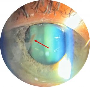
A young, short-sighted man rubs pigment loose as his iris chafes against the lens zonules, and that pigment quietly clogs the drainage meshwork.
By the time the pressure climbs, the tell-tale signs — a vertical smudge on the cornea, a jet-black angle, and spoke-like iris defects — have been sitting there for years.
For clinicians, recognizing Pigmentary Glaucoma matters because it strikes a younger, myopic population and can be aggressive if the pressure spikes are missed.
The whole story can be read at the slit lamp, from the cornea to the iris to the angle.
What Is Pigmentary Glaucoma?
Pigmentary Glaucoma is a secondary open-angle glaucoma that develops on the background of pigment dispersion syndrome.
Its defining chain is:
- Pigment dispersion — iris pigment liberated by chafing against the lens zonules
- Deposition of that pigment throughout the anterior segment
- Accumulation of pigment in the trabecular meshwork, impairing outflow
- A rise in intraocular pressure and glaucomatous optic nerve damage
Pigment dispersion is the syndrome; pigmentary glaucoma is what happens when the pressure follows.
Epidemiology and Mechanism
The demographic is distinctive.
- Typically young to middle-aged, myopic men
- A concave peripheral iris allows contact between the iris and the lens zonules
- This reverse pupillary block configuration drives the mechanical rubbing that releases pigment
- Exercise or pupillary dilation can trigger a shower of pigment and a pressure spike
The mechanical chafing of iris against zonule is the engine of the whole process.
All-fit Slit-Lamp Adapter
Record and share exactly what you see at the slit lamp. One adapter fits any slit lamp or surgical microscope — and any smartphone.
From Choroida — the team behind this siteClinical Presentation
Symptoms
- Often asymptomatic until the glaucoma is advanced
- Intermittent blurring, halos, or eye ache after exercise, from pigment-induced pressure spikes
Examination Findings
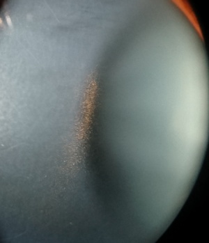
- A Krukenberg spindle — a vertical band of pigment on the corneal endothelium
- Heavy, homogeneous trabecular meshwork pigmentation on gonioscopy
- Mid-peripheral, spoke-like iris transillumination defects
- Pigment on the lens surface and zonules, and a deep anterior chamber
A Krukenberg spindle with spoke-like iris transillumination in a myopic young adult is the classic triad.
Diagnostic Evaluation
The assessment documents the pigment and stages the glaucoma.
- Slit-lamp examination for the Krukenberg spindle and iris transillumination
- Gonioscopy to grade the trabecular pigmentation and confirm an open angle
- Intraocular pressure measurement, including after exercise if spikes are suspected
- Optic nerve imaging and visual fields to detect and monitor glaucomatous damage
Transillumination and a heavily pigmented open angle separate pigmentary glaucoma from other secondary glaucomas.
Differential Diagnosis
Other causes of a pigmented angle and secondary glaucoma include:
- Pseudoexfoliation glaucoma — flaky material on the lens and pupil margin, with patchy angle pigment
- Uveitis with pigment release — accompanied by inflammation
- Post-traumatic angle recession — a history of injury and angle changes
- Pigment dispersion without glaucoma — the same signs but a normal pressure and nerve
The young myopic patient, the spindle, and the spoke-like transillumination point specifically to the pigmentary mechanism.
Management
Treatment lowers the pressure and addresses the mechanism where possible.
- Pressure-lowering medication as first-line therapy, as in other open-angle glaucomas
- Laser trabeculoplasty is often effective in these heavily pigmented angles, though pressure spikes must be watched for
- Laser peripheral iridotomy can flatten the concave iris and reduce iridozonular contact in selected cases
- Surgery for pressures not controlled medically or with laser
Because it affects younger eyes over many decades, sustained pressure control is especially important.
Prognosis
The course varies but is often manageable.
- Pigment release and pressure may lessen with age as the iris configuration changes
- Well-controlled disease carries a good long-term prognosis
- Undetected pressure spikes and poor control are the main threats to the optic nerve
In practice, recognising pigment dispersion early lets you watch for, and treat, the glaucoma before the nerve is damaged.
Would you like to document anterior segment pigment findings with your smartphone?


Document what you see
Two smartphone imaging tools built for everyday clinic use — one for the slit lamp, one for the fundus.
From Choroida — the team behind this siteReferences
- Sugar HS, Barbour FA. “Pigmentary Glaucoma: A Rare Clinical Entity.” American Journal of Ophthalmology. 1949;32(1):90–92.
- Niyadurupola N, Broadway DC. “Pigment Dispersion Syndrome and Pigmentary Glaucoma: A Major Review.” Clinical & Experimental Ophthalmology. 2008;36(9):868–882.
- Scheie HG, Cameron JD. “Pigment Dispersion Syndrome: A Clinical Study.” British Journal of Ophthalmology. 1981;65(4):264–269.
- American Academy of Ophthalmology. “Pigment Dispersion Syndrome and Pigmentary Glaucoma.” EyeWiki.
Test yourself
A few questions straight from this article.
-
Pigment dispersion syndrome is said to have become pigmentary glaucoma when what occurs?
Pigment dispersion is the syndrome; pigmentary glaucoma is diagnosed once meshwork obstruction raises pressure and glaucomatous optic nerve damage follows. -
What mechanism liberates iris pigment in pigment dispersion syndrome?
A concave peripheral iris lets the iris rub against the lens zonules in a reverse pupillary block configuration, and that mechanical chafing releases the pigment. -
Which patient group is typically affected by pigmentary glaucoma?
The demographic is distinctive, and it matters: affected eyes are young, so they must be kept under control for many decades. -
What is a Krukenberg spindle in pigment dispersion syndrome?
Liberated pigment is deposited in a vertical band on the corneal endothelium, giving the tell-tale smudge visible at the slit lamp. -
What pattern do the iris transillumination defects take in pigment dispersion syndrome?
Pigment lost where the iris rubs the zonules leaves mid-peripheral spoke-like defects, which with a Krukenberg spindle in a myopic young adult form the classic triad. -
What can trigger a shower of pigment and a pressure spike in pigment dispersion syndrome?
Exercise or dilation can release a burst of pigment and spike the pressure, which is why pressure may be checked after exercise when spikes are suspected. -
Which finding suggests pseudoexfoliation glaucoma rather than pigmentary glaucoma?
Pseudoexfoliation deposits flaky material on the lens and pupil margin and pigments the angle patchily, rather than the even heavy pigmentation of pigment dispersion. -
What is the first-line treatment for pigmentary glaucoma?
As in other open-angle glaucomas, medication comes first; laser trabeculoplasty and surgery follow when pressure is not controlled. -
Why is laser peripheral iridotomy considered in selected patients with pigmentary glaucoma?
Iridotomy addresses the mechanism itself by flattening the backward-bowed iris so it no longer rubs the zonules and sheds pigment. -
How does pigment release typically behave over time in pigment dispersion syndrome?
Pigment release and pressure often settle as the iris configuration changes with age, and well-controlled disease carries a good long-term prognosis.
