
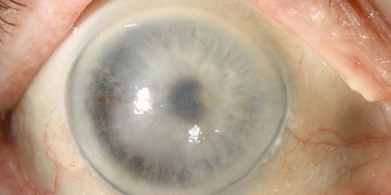
Descemet membrane detachment (DMD) is a separation of Descemet’s membrane and the corneal endothelium from the overlying stroma, most often occurring as a complication of intraocular surgery — cataract surgery above all — though it can also follow trauma or, rarely, occur spontaneously.
Because Descemet’s membrane and its endothelium are what actively pump fluid out of the cornea to keep it clear, a detachment that goes unrecognized or untreated leads to localized or diffuse corneal edema that can persist long after an otherwise uncomplicated surgery, undermining what would otherwise have been an unremarkable postoperative course.
Mechanism
During cataract surgery, instruments or irrigating fluid entering through the clear corneal incision can shear Descemet’s membrane away from the overlying stroma at the incision site, creating a small flap or, in more extensive cases, a larger detachment that can extend across a significant portion of the cornea.
Because Descemet’s membrane is elastic and tends to curl, a detached segment often rolls up on itself (a “Descemet’s membrane roll”) rather than lying flat, which is part of what makes small detachments visible on careful slit-lamp exam even when subtle.
This tendency to roll rather than fold flat is also part of why extending gas tamponade to cover the entire detached segment matters — a partially treated roll can persist folded even when the flatter, untreated edge might otherwise have reattached spontaneously.
Risk Factors
- Blunt or dull surgical instruments, which increase shearing force during insertion
- Incorrect instrument orientation on entry into the anterior chamber
- Shallow anterior chamber during surgery
- Pre-existing corneal endothelial compromise (Fuchs endothelial dystrophy, prior corneal surgery)
- High-pressure irrigation directed inadvertently beneath Descemet’s membrane rather than into the anterior chamber proper
All-fit Slit-Lamp Adapter
Record and share exactly what you see at the slit lamp. One adapter fits any slit lamp or surgical microscope — and any smartphone.
From Choroida — the team behind this siteClinical Presentation
Small, localized detachments near the surgical incision are often asymptomatic and self-limited, resolving spontaneously as the membrane reattaches over the following days to weeks.
Larger detachments present with corneal edema and reduced vision that is disproportionate to what would be expected from routine postoperative inflammation, and the edema classically corresponds precisely to the area where Descemet’s membrane has separated, sparing the rest of the cornea where the membrane remains attached — a pattern that is itself a useful diagnostic clue.
Exam and Imaging Findings
- Localized corneal edema, often near the surgical wound, with a sharp border corresponding to the edge of the detachment
- A visible membrane or scroll within the anterior chamber in more significant detachments, sometimes seen floating free of the posterior stroma on careful slit-lamp exam
- Anterior segment OCT is the most useful confirmatory tool, directly visualizing the separation between Descemet’s membrane and the overlying stroma, and allowing measurement of detachment height and extent
- Ultrasound biomicroscopy as an alternative when OCT is unavailable or when the view is obscured by significant corneal edema
Differential Diagnosis
- Ordinary postoperative corneal edema — diffuse rather than sharply localized, gradually improving over the expected postoperative timeline
- Endothelial decompensation from pre-existing Fuchs dystrophy, unmasked or worsened by surgery — typically more diffuse and without a visible detached membrane on imaging
- Descemet’s membrane tear (as opposed to detachment) — a discontinuity rather than a separation, sometimes seen in the context of forceps trauma at birth or acute hydrops
Grading and Management
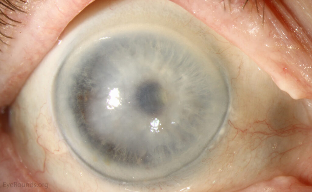
Several grading schemes exist, generally based on the height of the detachment relative to the corneal thickness and the extent of the visual axis involved, and while none has become universally standardized, the practical distinction that matters most is simply whether the detachment is small and peripheral or large and central.
Small, low-lying detachments not involving the visual axis can be observed, because a meaningful proportion resolve spontaneously as normal endothelial pump function gradually reattaches the membrane over days to a few weeks, without needing any active intervention.
Larger detachments, or those involving the visual axis with significant corneal edema, are treated with an intracameral gas (usually air or a short-acting gas such as SF6) injection — a “pneumo-descemetopexy” — which tamponades the detached membrane against the posterior stroma, allowing it to reattach while the endothelial pump re-establishes normal corneal deturgescence.
Persistent detachments unresponsive to gas tamponade, when extensive or associated with significant endothelial cell loss, may ultimately require endothelial keratoplasty if the cornea fails to clear on its own, though this is reserved for cases where conservative management has genuinely failed rather than being a first-line approach, given the additional surgical risk and recovery time a corneal transplant entails.
Prognosis
Most DMDs identified and treated promptly resolve with good visual recovery, when the detachment is recognized early and the underlying endothelium was healthy before surgery.
Outcomes are less favorable when the detachment is extensive, longstanding before treatment, or occurs in an eye with pre-existing endothelial compromise, because the endothelial cell reserve needed to re-establish normal corneal clarity may already be limited.
This is why prompt recognition in the early postoperative period matters so much — a detachment identified and treated within the first week or two generally has a meaningfully better prognosis than one that goes unnoticed until corneal edema has already been present for an extended period.


Document what you see
Two smartphone imaging tools built for everyday clinic use — one for the slit lamp, one for the fundus.
From Choroida — the team behind this siteReferences
- Jain R, Assi A, Murthy S, Vajpayee RB. Descemet’s membrane detachment. In: Cornea and External Eye Disease.
- Kim T, Hasan SA. A new technique for repairing Descemet membrane detachments using intracamerally injected air. Archives of Ophthalmology.
- Sharma N, Maharana P, Singh G, Titiyal JS. Pneumatic descemetopexy for the management of descemet membrane detachment. Cornea.
- American Academy of Ophthalmology. Basic and Clinical Science Course, Section 8: External Disease and Cornea.
Test yourself
A few questions straight from this article.
-
What anatomical event defines Descemet membrane detachment?
Descemet membrane detachment is a separation of Descemet's membrane together with the corneal endothelium away from the overlying corneal stroma. -
Why does Descemet membrane detachment produce corneal edema?
Descemet's membrane and its endothelium actively pump fluid out of the cornea; once detached, that deturgescence fails and the cornea becomes edematous. -
Which intraoperative event most often causes Descemet membrane detachment in cataract surgery?
Instruments or irrigating fluid passing through the clear corneal incision can shear Descemet's membrane off the stroma at the wound, creating a flap or a wider detachment. -
Which surgical factor raises the risk of Descemet membrane detachment during cataract surgery?
Blunt or dull instruments increase shearing force on entry. A shallow chamber, wrong instrument orientation and high-pressure irrigation beneath the membrane add further risk. -
Which corneal edema pattern suggests Descemet membrane detachment rather than ordinary postoperative edema?
The edema corresponds precisely to the detached segment and spares cornea where the membrane is still attached, whereas routine postoperative edema is diffuse. -
Which imaging tool best confirms a suspected Descemet membrane detachment?
Anterior segment OCT directly shows the separation between Descemet's membrane and stroma and lets you measure detachment height and extent. -
What distinguishes a Descemet's membrane tear from a Descemet membrane detachment?
A tear is a break in the membrane's continuity, classically after birth forceps trauma or acute hydrops, while detachment is separation of an intact membrane from stroma. -
Which Descemet membrane detachment can reasonably be observed after cataract surgery?
Small peripheral detachments are often asymptomatic and a meaningful proportion reattach spontaneously over days to a few weeks as endothelial pump function returns. -
How is a large Descemet membrane detachment involving the visual axis usually treated?
Air or a short-acting gas such as SF6 is injected to tamponade the membrane against posterior stroma so it can reattach and the endothelial pump can clear the cornea. -
Which feature predicts a poorer visual outcome after Descemet membrane detachment?
Outcomes are worse when the detachment is extensive or longstanding, or when endothelial reserve was already limited before surgery, since clearing the cornea depends on that reserve.
