
In a highly myopic eye, a small, round, dark pigmented patch sometimes sits right at the fovea.
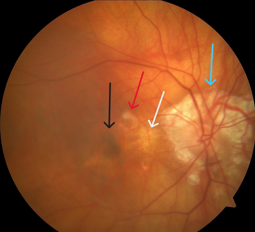
It is slightly raised, sharply bordered, and has a pale rim around it.
That is a Fuchs or Forster-Fuchs spot, and it is the footprint of an old myopic choroidal neovascular membrane.
How it forms
Myopic CNV tends to be small and flat.
After it leaks or bleeds, it often regresses, and the RPE responds by proliferating over the scar.
The pigment accumulates and a hyperpigmented plaque results.
The surrounding atrophy gives the characteristic pale halo, which may enlarge over the years.
Typical features
- Occurs in eyes with axial length usually above 26 to 27 mm
- Often found in patients in the third to sixth decades
- May follow a period of sudden central blur or metamorphopsia that the patient barely remembers
- Associated with lacquer cracks, posterior staphyloma, and chorioretinal atrophy (see pathologic myopia META-PM classification)
Fundus Explorer Pro
Photograph the retinal findings described here with the phone already in your pocket — 22 D optics and built-in illumination in one handheld unit.
From Choroida — the team behind this siteWhy it matters
The spot itself does not need treatment.
It does tell you the patient has already had CNV in that eye and may have it again, in the same eye or the fellow eye.
Fresh CNV beside an old spot presents with a small hemorrhage, new distortion, or a change in the lesion’s outline (see myopic choroidal neovascularization).
Differential diagnosis
- Choroidal nevus, which is not surrounded by atrophy and shows no lacquer cracks
- Hemorrhage, which fades in weeks, whereas a Fuchs spot is permanent
- Combined hamartoma and CHRPE, which are in different locations and settings
- Disciform scars of age-related CNV, which occur in older patients without high myopia
Natural history
Many Fuchs spots appear after a small, unnoticed episode.
In the Pathologic Myopia classification, the lesion is a macular atrophy-related finding.
Over years, the patch of pigment may shrink slightly, while the halo of chorioretinal atrophy tends to expand.
Patients occasionally describe a “fixed dark spot” that has been present for as long as they can remember.
When vision falls substantially, the cause is often atrophy at the fovea, not a new membrane, which is why OCT is important to decide whether anti-VEGF treatment has anything to offer.
Examination points
- Look for a hemorrhage at the edge of the spot, since that shows reactivation
- Check the fellow eye, which carries a significant risk of developing CNV in highly myopic patients
- Measure axial length when available, because it affects risk estimates and counselling
- Add a note of other myopic complications such as peripapillary atrophy, tilted disc and staphyloma
Management
- Document the lesion with color photographs and OCT, which will show hyperreflective RPE with variable subretinal fibrosis
- Give an Amsler grid and instruct the patient to report any new distortion
- Treat new active CNV with anti-VEGF injections, usually with a short course
How to separate it from a fresh membrane
A new myopic CNV is usually small, gray or yellow-green, and may be surrounded by a thin ring of blood. On OCT it is a small hyperreflective lesion above the RPE with minimal fluid. A Fuchs spot, by contrast, is dark, stable, pigmented, and has a pale atrophic halo, so a change in color or the appearance of blood at its edge is the clue to reactivation.
Counselling highly myopic patients
Patients with a Fuchs spot should know that they are at risk of further membranes and of progressive macular atrophy. They should be taught to check each eye separately with an Amsler grid, to report any new distortion or blur at once, and to avoid delaying a visit because the other eye is "good". Because myopic CNV responds well to a small number of anti-VEGF injections, early presentation preserves the most vision.
Where available, long-term follow-up with OCT every six to twelve months is a reasonable standard.
Imaging the myopic macula
Optical coherence tomography shows the Fuchs spot as a hyperreflective, thickened RPE complex with variable subretinal fibrosis and thinning of the surrounding choroid. Fundus autofluorescence shows a dark center with a hyperautofluorescent rim in some cases, and a wide area of reduced signal where atrophy has developed. Widefield imaging helps to document the extent of staphyloma and peripheral changes. These images are useful baselines, since progression of atrophy is slow and best judged by comparison.
OCT angiography can show a small vascular complex within an active lesion, but it is not needed in routine care.
Prognosis
Central acuity is limited by the scar and atrophy.
The long-term risk is progressive foveal atrophy, which is why myopic maculopathy is a leading cause of irreversible vision loss in some populations.


Document what you see
Two smartphone imaging tools built for everyday clinic use — one for the slit lamp, one for the fundus.
From Choroida — the team behind this siteReferences
- Avila MP, Weiter JJ, Jalkh AE, Trempe CL, Pruett RC, Schepens CL. Natural history of choroidal neovascularization in degenerative myopia. Ophthalmology. 1984;91:1573-1581.
- Ohno-Matsui K, Kawasaki R, Jonas JB, et al. International photographic classification and grading system for myopic maculopathy. Am J Ophthalmol. 2015;159:877-883.
- Wong TY, Ohno-Matsui K, Leveziel N, et al. Myopic choroidal neovascularisation: current concepts and update on clinical management. Br J Ophthalmol. 2015;99:289-296.
- Ohno-Matsui K, Lai TY, Lai CC, Cheung CM. Updates of pathologic myopia. Prog Retin Eye Res. 2016;52:156-187.
