
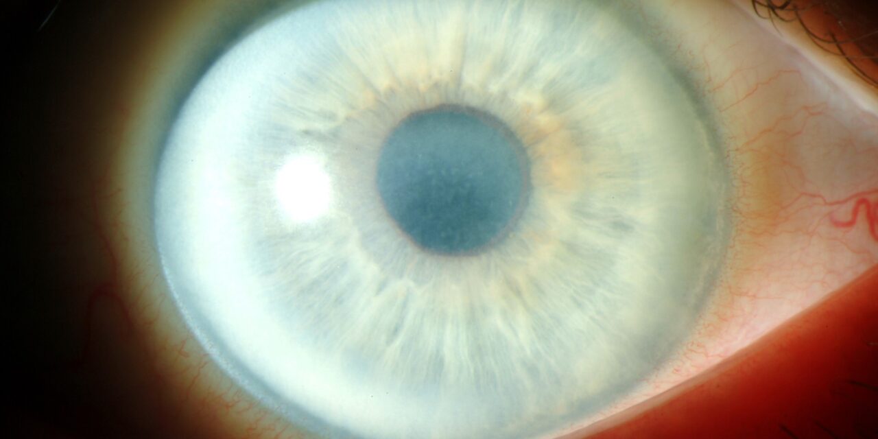
Schnyder corneal dystrophy is a rare, autosomal dominant stromal dystrophy caused by abnormal deposition of cholesterol and phospholipid within the corneal stroma, and it is genuinely notable for being one of the few corneal dystrophies with a well-established, clinically relevant systemic association: a substantial proportion of affected patients also have systemic hyperlipidemia.
Recognizing the corneal finding should prompt a lipid panel and broader cardiovascular risk assessment rather than being treated as a purely isolated ocular curiosity.
This makes Schnyder dystrophy a useful reminder that even a rare, genetically determined corneal finding can carry genuine, actionable systemic implications, and that a dedicated eye exam sometimes surfaces information relevant well beyond the visual system.
Genetics and Systemic Association
Schnyder corneal dystrophy results from mutations in UBIAD1, a gene involved in cholesterol and vitamin K2 metabolism, which explains both the corneal lipid deposition characteristic of the disease and its recognized, though not universal, association with systemic dyslipidemia.
While not every patient with Schnyder dystrophy has abnormal systemic lipids, the association is strong enough, and simple enough to screen for, that a fasting lipid panel is a reasonable and low-cost part of the initial workup once the corneal diagnosis is made or suspected.
Clinical Presentation and Staging
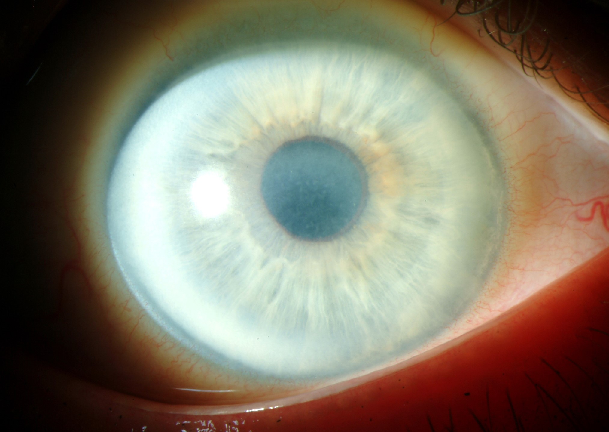
The classic, though not universally present, early finding is a central, ring-shaped or disc-shaped area of fine, glistening, needle-like crystals within the anterior stroma, sometimes accompanied by a surrounding arcus-like ring of lipid deposition at the corneal periphery — reminiscent of, but distinct from, ordinary arcus senilis, discussed in this site’s general coverage of that finding, given its earlier onset and association with the central, crystalline stromal changes rather than occurring as an isolated peripheral finding.
Not every patient shows visible crystals, however: a meaningful subset of cases present instead with more diffuse stromal haze without the classic crystalline pattern, which can make the diagnosis less immediately obvious without a high index of suspicion in a patient with a relevant family history.
All-fit Slit-Lamp Adapter
Record and share exactly what you see at the slit lamp. One adapter fits any slit lamp or surgical microscope — and any smartphone.
From Choroida — the team behind this siteProgression
The disease is slowly progressive over decades, with stromal haze and clouding gradually increasing and progressively affecting vision, generally becoming visually significant only in mid-to-late adulthood despite the underlying deposition process beginning much earlier in life.
Corneal sensation is often reduced in longstanding disease, an additional finding that, while not unique to Schnyder dystrophy, adds to the overall clinical picture in more advanced cases.
Exam Findings
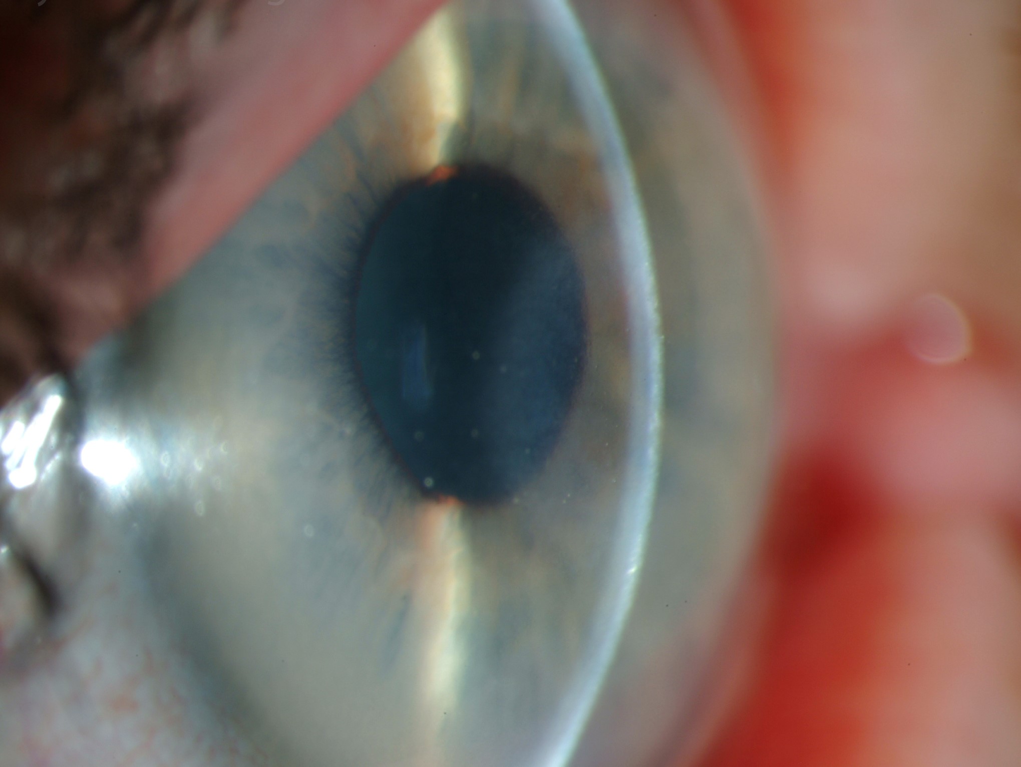
- Central, ring or disc-shaped, fine, needle-like or glistening crystalline deposits in the anterior-to-mid stroma, when present — genuinely classic and highly suggestive when clearly visible, though absent in a meaningful proportion of cases
- A peripheral arcus-like ring of lipid deposition, sometimes present at a notably younger age rather than typical arcus senilis would be expected
- Progressive, diffuse stromal haze, present in essentially all cases regardless of whether the classic crystalline pattern is visible
- Reduced corneal sensation in more advanced, longstanding disease
- Bilateral, generally symmetric involvement, consistent with the autosomal dominant inheritance pattern
Differential Diagnosis
- Arcus senilis — an isolated peripheral lipid ring without central crystalline deposits or progressive central stromal haze, and typically occurring at an older age rather than the arcus-like ring sometimes seen in Schnyder dystrophy, discussed in this site’s general coverage of that finding
- Bietti crystalline dystrophy — a different, retinal (rather than corneal) crystalline deposition disorder, though occasional corneal crystals have also been reported in that condition, making a careful distinction between the two based on the primary site and pattern of crystal deposition important when the diagnosis is uncertain
- Other stromal corneal dystrophies (lattice, granular, macular) — each has its own distinct deposit pattern and composition, discussed in their own dedicated articles on this site, generally distinguishable from Schnyder dystrophy’s characteristic central crystalline or diffuse haze pattern combined with its systemic lipid association
- Cystinosis — also causes corneal crystal deposition, but of cystine rather than cholesterol/lipid, and accompanied by other systemic findings (renal disease, growth failure) that are absent in isolated Schnyder dystrophy, discussed in this site’s coverage of ocular cystinosis
Management
Mild, early disease requires no ocular treatment beyond monitoring, since the primary goal at this stage is simply tracking progression over time.
Identifying and treating any underlying systemic dyslipidemia is worthwhile both for its own cardiovascular health implications and, potentially, as a general health measure independent of any direct effect on corneal disease progression, though evidence that treating systemic lipids specifically slows corneal deposition is limited, so this recommendation is made primarily for general cardiovascular benefit rather than expected corneal improvement.
As stromal haze progressively affects vision, phototherapeutic keratectomy can improve clarity for more superficial deposits, though — similar to the other stromal dystrophies discussed elsewhere on this site — some degree of recurrence over subsequent years is expected given that the underlying metabolic deposition process continues.
Corneal transplantation (lamellar or penetrating, depending on the depth and extent of stromal involvement) is reserved for more advanced disease with significant visual impact, and patients should be counseled, as with other stromal dystrophies, that the same underlying process can eventually affect a graft over subsequent years, because transplantation replaces the diseased tissue without altering the patient’s ongoing metabolic tendency to deposit lipid within corneal stroma.


Document what you see
Two smartphone imaging tools built for everyday clinic use — one for the slit lamp, one for the fundus.
From Choroida — the team behind this siteReferences
- Weiss JS. Schnyder corneal dystrophy. Current Opinion in Ophthalmology.
- Orr A, Dubé MP, Marcadier J, et al. Mutations in the UBIAD1 gene, encoding a potential prenyltransferase, are causal for Schnyder corneal dystrophy. PLoS One.
- American Academy of Ophthalmology. Basic and Clinical Science Course, Section 8: External Disease and Cornea.
