
An eyelid coloboma is a congenital, full-thickness defect in the eyelid, most often the upper lid, and because even a modest defect can leave the underlying cornea chronically exposed in a newborn who cannot yet protect their own eye through normal blink and Bell’s phenomenon reflexes as robustly as an older child, this is a condition where the timeline for repair is often measured in days, not months.
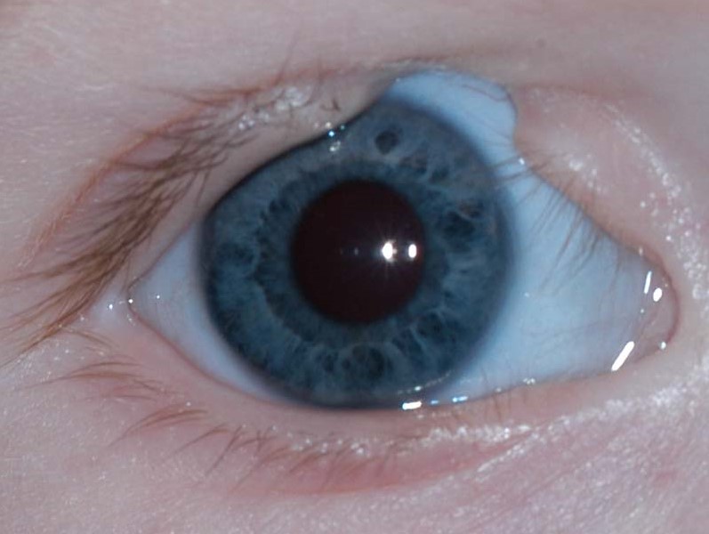
What an Eyelid Coloboma Is
Eyelid coloboma results from incomplete fusion of the developing eyelid tissue during gestation, producing a notch or gap, typically triangular or wedge-shaped, involving the full thickness of the eyelid: skin, muscle, tarsus, and conjunctiva.
The upper eyelid, particularly its medial to central portion, is affected more often than the lower eyelid, and the size of the defect varies considerably, from a small notch to a defect encompassing a substantial portion of the lid.
Associated Conditions
Eyelid coloboma can occur as an isolated finding or as part of a broader craniofacial syndrome, and recognizing this association is important for appropriate systemic evaluation.
- Goldenhar syndrome (oculo-auriculo-vertebral spectrum), frequently associated with upper eyelid coloboma along with other craniofacial and vertebral anomalies
- Treacher Collins syndrome, more classically associated with lower eyelid coloboma
- Amniotic band syndrome, in which constricting fibrous bands can cause eyelid and other congenital defects
- Frontonasal dysplasia and other craniofacial clefting syndromes, including Tessier facial cleft patterns
- Isolated, non-syndromic eyelid coloboma also occurs
All-fit Slit-Lamp Adapter
Record and share exactly what you see at the slit lamp. One adapter fits any slit lamp or surgical microscope — and any smartphone.
From Choroida — the team behind this siteWhy Prompt Assessment and Management Matter
A full-thickness eyelid defect leaves the underlying cornea inadequately protected, since normal blinking and lid closure, essential for spreading tear film and protecting the ocular surface, cannot occur properly across the gap.
In a neonate, this exposure risk is compounded by generally less robust Bell’s phenomenon (the protective upward eye rotation during attempted lid closure) than is typical in older children and adults, so even a modest coloboma can lead rapidly to exposure keratopathy, corneal ulceration, and, in severe untreated cases, corneal perforation and loss of the eye.
Initial Management
- Immediate lubrication with preservative-free ointment and, when needed, a moisture chamber, to protect the exposed cornea while definitive surgical planning proceeds
- Careful assessment of the cornea for any early exposure changes, requiring close, frequent monitoring in the initial period after birth
- Evaluation for associated craniofacial or systemic anomalies, given the recognized syndromic associations, coordinated with genetics and craniofacial specialists as appropriate
Timing and Goals of Surgical Repair
Small colobomas with minimal corneal exposure risk may be observed initially with aggressive lubrication, but larger defects, or any defect associated with early signs of corneal exposure, require urgent surgical closure, sometimes within the first days of life, to protect the cornea before permanent damage occurs.
The primary goal of early repair is protective: restoring adequate lid closure and corneal coverage, with attention to eyelid contour and cosmesis balanced against the more urgent priority of preventing corneal injury.
Surgical Techniques
- Direct closure: for smaller defects, primary closure of the coloboma edges, in anatomic layers (conjunctiva, tarsus, muscle, skin), can achieve adequate closure without needing additional tissue
- Local flaps: for larger defects that cannot be closed directly without excessive tension, local tissue flaps, such as a Tenzel semicircular flap or similar techniques adapted from eyelid reconstruction more broadly, can be used to recruit adjacent tissue
- Staged reconstruction: very large defects may require a staged approach, with an initial procedure to achieve safe corneal coverage followed by later revision surgery, once the child is older, to optimize eyelid contour and function
Long-Term Follow-Up
Children undergo ongoing follow-up to monitor eyelid position and function as they grow, since growth can alter the appearance and function of the initial repair, and secondary revision surgery is not uncommon, particularly for larger initial defects, to improve eyelid contour, lash line appearance, or residual lagophthalmos.
Prognosis
With prompt recognition and timely surgical repair before significant corneal exposure damage occurs, visual outcomes are generally good, and the eyelid can be reconstructed to provide adequate long-term corneal protection.
Delayed recognition or treatment, allowing corneal exposure to progress, is the main driver of poor visual outcomes in this condition, which is why early, sometimes urgent, involvement of both pediatric ophthalmology and oculoplastic surgery is important as soon as an eyelid coloboma is identified.


Document what you see
Two smartphone imaging tools built for everyday clinic use — one for the slit lamp, one for the fundus.
From Choroida — the team behind this siteReferences
- Bowling B. Kanski’s Clinical Ophthalmology: A Systematic Approach. 8th ed. Edinburgh: Elsevier; 2016.
- Meyer DR, Fincham T. Congenital eyelid colobomas: pathogenesis and management. Ophthalmic Plast Reconstr Surg. 2003;19:212-217.
- Tenzel RR, Stewart WB. Eyelid reconstruction by the semicircle flap technique. Ophthalmology. 1978;85:1164-1169.
- Baijens LW, Freihofer HP. Characteristics and surgical treatment of the Treacher-Collins syndrome. J Craniomaxillofac Surg. 2000;28:91-98.
