
Foville syndrome is a brainstem stroke syndrome caused by a lesion in the dorsal pons, producing a combination of ipsilateral sixth and seventh cranial nerve palsy, a horizontal gaze palsy, and contralateral body weakness or sensory loss.
It belongs to a family of eponymous brainstem syndromes defined by the specific combination of cranial nerve findings on the side of the lesion paired with long-tract findings on the opposite side, a pattern called crossed or alternating brainstem signs.
Recognizing this crossed pattern is what allows a clinician to localize a lesion to the brainstem, and specifically to the pons, purely from the bedside exam, well before any imaging is obtained.
Foville syndrome is one of several related pontine syndromes with overlapping but distinguishable findings, and precise localization within this family depends on exactly which structures a given lesion happens to involve.
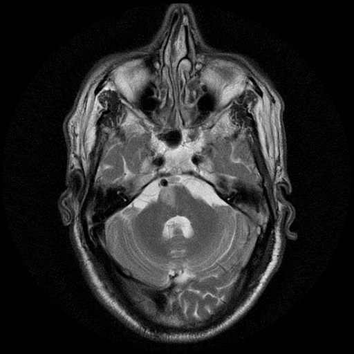
Anatomy and Localization
The dorsal pons houses the abducens nucleus, the facial nerve as it wraps around the abducens nucleus in a loop called the facial colliculus, and the paramedian pontine reticular formation, the center responsible for coordinating horizontal conjugate gaze toward the same side.
A lesion in this region, most often from occlusion of a paramedian branch of the basilar artery, damages all of these structures together, which is why the syndrome combines a sixth nerve palsy, a seventh nerve palsy, and a horizontal gaze palsy rather than any one of these findings in isolation.
The corticospinal tract, still ascending through the pons before it decussates lower in the medulla, is also affected, producing contralateral weakness, which is what gives Foville syndrome its characteristic crossed pattern of ipsilateral cranial nerve findings with contralateral body weakness.
The exact combination of findings in a given patient can vary somewhat depending on precisely how far the lesion extends within the dorsal pons, which is why textbook descriptions of the syndrome sometimes differ slightly from one another.
Clinical Findings
- Ipsilateral sixth nerve palsy, causing an inability to abduct the eye on the side of the lesion
- Ipsilateral horizontal gaze palsy, an inability to look toward the side of the lesion with either eye, reflecting involvement of the gaze center rather than the sixth nerve alone
- Ipsilateral facial weakness from seventh nerve involvement, affecting both the upper and lower face given the lesion’s location within the facial nerve’s course rather than distal to it
- Contralateral hemiparesis or hemisensory loss from corticospinal or, less consistently, sensory tract involvement
All-fit Slit-Lamp Adapter
Record and share exactly what you see at the slit lamp. One adapter fits any slit lamp or surgical microscope — and any smartphone.
From Choroida — the team behind this siteDistinguishing Gaze Palsy From Isolated Sixth Nerve Palsy
An isolated sixth nerve palsy limits abduction of one eye only, while a horizontal gaze palsy prevents both eyes from moving conjugately toward the affected side, a distinction made by testing each eye individually versus testing conjugate gaze.
This distinction matters for localization: an isolated sixth nerve palsy points toward a lesion of the nerve itself, anywhere along its long intracranial course, while a true gaze palsy specifically implicates the pontine gaze center and strongly supports a brainstem, rather than a peripheral nerve, localization.
Recognizing a true gaze palsy on exam is, in practice, one of the more useful bedside clues for suspecting a brainstem process before imaging confirms it.
Causes
Ischemic stroke from small-vessel or paramedian branch occlusion of the basilar artery is the classic and most common cause, particularly in a patient with vascular risk factors.
Demyelinating disease, a brainstem tumor, or, less commonly, hemorrhage can produce a similar clinical picture through involvement of the same anatomic structures.
The acuity of onset is a useful clue for prioritizing the differential: a sudden onset favors stroke, while a more gradual course raises the possibility of a tumor or demyelinating process instead.
Evaluation and Management
Urgent neuroimaging, with MRI preferred over CT for its superior sensitivity to brainstem pathology, is warranted in any patient presenting with this pattern of findings, following standard acute stroke protocols when the presentation and timeline are consistent with an ischemic event.
Management follows the underlying cause: acute stroke management and secondary prevention for an ischemic cause, or the relevant treatment pathway for a demyelinating or neoplastic cause when imaging points elsewhere.
Coordination with neurology is essential both for the acute workup and for longer-term management and rehabilitation, since recovery of cranial nerve and motor function after a pontine stroke typically requires sustained multidisciplinary rehabilitation.
For the ophthalmologist specifically, the practical value of recognizing Foville syndrome lies in the gaze palsy and facial weakness findings, which can be the first clues that steer a patient toward urgent stroke evaluation rather than a peripheral ophthalmic workup.


Document what you see
Two smartphone imaging tools built for everyday clinic use — one for the slit lamp, one for the fundus.
From Choroida — the team behind this siteReferences
- American Academy of Ophthalmology. Basic and Clinical Science Course, Section 5: Neuro-Ophthalmology.
- Liu GT, Volpe NJ, Galetta SL. Neuro-Ophthalmology: Diagnosis and Management.
- Kumral E, Bayulkem G, Evyapan D. Clinical spectrum of pontine infarction: clinical-MRI correlations. Journal of Neurology.
Test yourself
A few questions straight from this article.
-
A lesion in which region of the brainstem produces Foville syndrome?
Foville syndrome follows a dorsal pontine lesion, which damages the abducens nucleus, the facial nerve loop and the pontine gaze centre together. -
What does the crossed pattern of the eponymous brainstem syndromes describe?
Cranial nerve findings on the side of the lesion paired with long-tract findings on the opposite side are what localise a lesion to the brainstem at the bedside. -
What is the facial colliculus?
In the dorsal pons the facial nerve wraps around the abducens nucleus in a loop called the facial colliculus, which is why one lesion affects both nerves. -
What does the paramedian pontine reticular formation control?
The paramedian pontine reticular formation is the centre coordinating horizontal conjugate gaze toward its own side, so its loss gives a gaze palsy. -
Which vascular event is the classic cause of Foville syndrome?
Ischaemic stroke from small-vessel or paramedian branch occlusion of the basilar artery is the classic and commonest cause, especially with vascular risk factors. -
Which pattern of facial weakness occurs in Foville syndrome?
Because the lesion sits within the facial nerve's course rather than distal to it, both the upper and lower face are weak on the side of the lesion. -
How is a horizontal gaze palsy distinguished from an isolated sixth nerve palsy?
An isolated sixth nerve palsy limits abduction of one eye only, whereas a gaze palsy stops both eyes moving conjugately toward the affected side. -
Involvement of which tract produces the contralateral weakness in Foville syndrome?
The corticospinal tract runs through the pons and decussates lower down, so a pontine lesion produces weakness on the opposite side of the body. -
Which imaging study is preferred when Foville syndrome is suspected?
Urgent MRI is preferred over CT because it is considerably more sensitive to brainstem pathology. -
Which cause does a gradual onset of Foville syndrome findings suggest?
Sudden onset favours stroke, while a more gradual course raises the possibility of a brainstem tumour or a demyelinating process instead.

Writes and reviews the clinical articles on Educate. Every post is checked against peer-reviewed sources before it goes live.


