
Congenital stationary night blindness (CSNB) is a genetically heterogeneous group of inherited retinal conditions causing non-progressive night vision impairment from birth.
The word “stationary” is doing real clinical work in its name: unlike the great majority of other inherited retinal dystrophies discussed elsewhere on this site, CSNB does not progress over a patient’s lifetime.
This is both reassuring for long-term prognosis and a key diagnostic feature distinguishing it from progressive conditions that can otherwise present with a broadly similar early clinical picture.
Why Recognizing “Stationary” Matters
A child diagnosed with night blindness in infancy or early childhood, whose parents are naturally anxious about future vision loss, benefits enormously from an accurate distinction between a stationary condition like CSNB — where visual function, once established, remains stable for life — and a progressive rod-cone dystrophy such as retinitis pigmentosa, discussed in its own dedicated article on this site, which carries an entirely different, more concerning long-term trajectory.
Confirming stability, typically through serial ERG testing over time showing no significant change, is central to reassuring families and to correctly counseling about prognosis.
Classification
- Schubert-Bornschein type — the more commonly encountered form, further divided into “complete” and “incomplete” subtypes based on specific ERG waveform characteristics, associated with mutations affecting the signal transmission between photoreceptors and bipolar cells; this type is frequently, though not always, X-linked and associated with high myopia
- Riggs type — a rarer form with a different underlying defect localized more to the photoreceptors themselves rather than the downstream bipolar cell signaling pathway, generally producing a somewhat different ERG pattern
Distinguishing complete from incomplete Schubert-Bornschein CSNB, and this type from Riggs type, relies substantially on detailed ERG waveform analysis rather than clinical exam findings alone, because the fundus itself is often unremarkable across these different genetic and electrophysiologic subtypes.
Fundus Explorer Pro
Photograph the retinal findings described here with the phone already in your pocket — 22 D optics and built-in illumination in one handheld unit.
From Choroida — the team behind this siteClinical Presentation
- Night blindness present from birth or very early infancy, often the presenting complaint once a child is old enough for the difficulty navigating in dim light to become apparent to parents
- Reduced visual acuity in many, though not all, patients, generally mild to moderate rather than severe
- High myopia, a strong and characteristic association particularly with the X-linked Schubert-Bornschein form, sometimes prompting the initial ophthalmic evaluation independent of any specifically reported night vision complaint
- Nystagmus and strabismus in some affected children, related to the reduced visual acuity present from early in visual development
- Photophobia in some patients, though generally less prominent than in cone-predominant conditions such as achromatopsia or cone-rod dystrophy, both discussed in their own dedicated articles on this site
Exam Findings
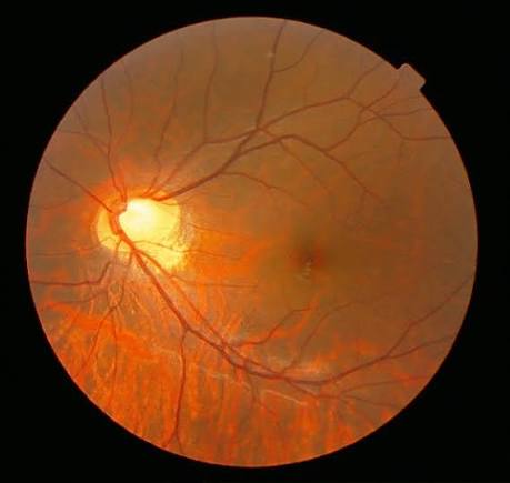
The fundus is often entirely normal in appearance, which — similar to the pattern discussed for Leber congenital amaurosis in this site’s dedicated article on that condition — means a normal-looking retina does not exclude the diagnosis and should not delay ERG testing in a child with unexplained night blindness.
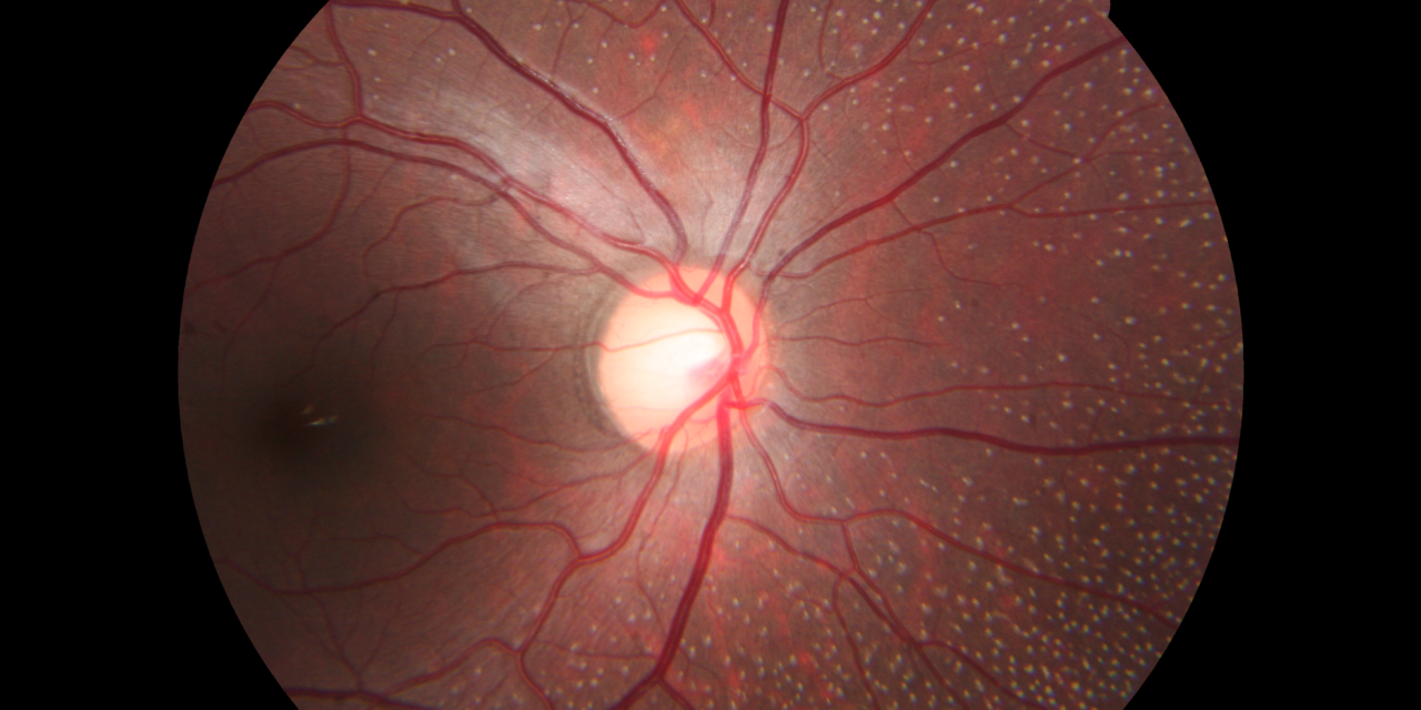
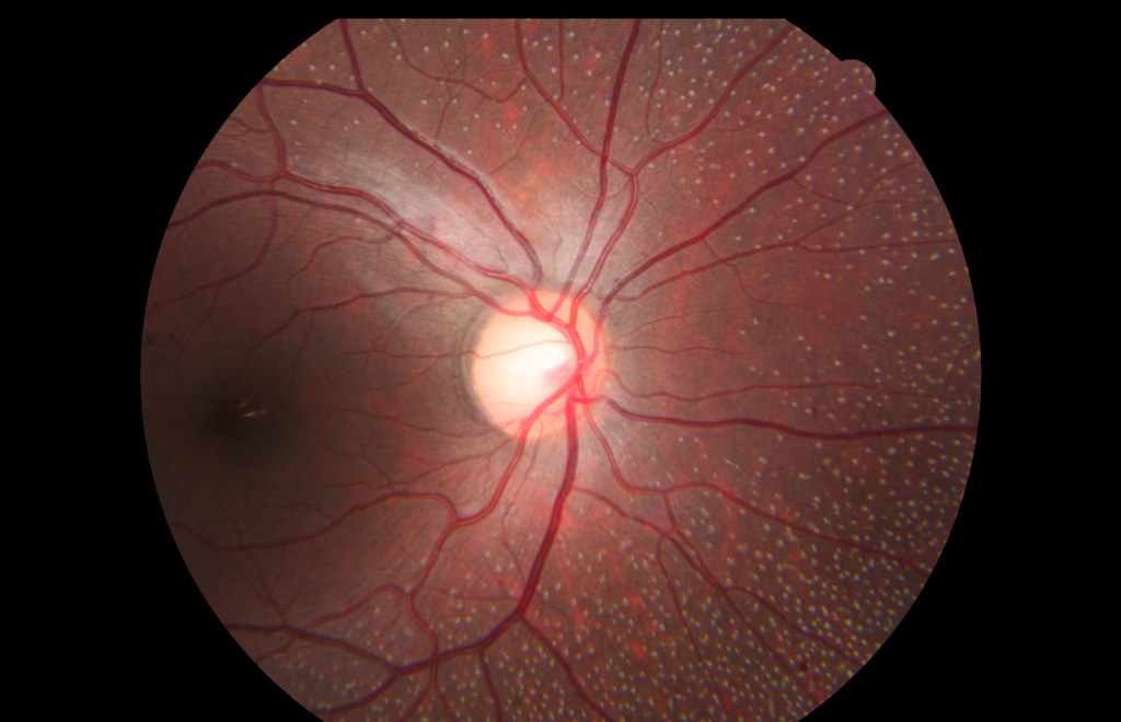
A notable variant, fundus albipunctatus, discussed in its own dedicated article on this site, does show characteristic small white retinal dots and represents a specific, visually distinct subtype within the broader CSNB spectrum related to delayed rather than absent rod photopigment regeneration.
Diagnostic Evaluation
Electroretinography is the essential diagnostic test.
The specific waveform pattern — the relationship between the a-wave and b-wave amplitudes under dark-adapted (scotopic) conditions — both confirms the diagnosis and helps distinguish the specific CSNB subtype, information that has genuine value for genetic counseling even though it does not change the overall favorable, stable prognosis shared across the CSNB spectrum.
Refraction is essential given the strong myopia association, particularly for the X-linked Schubert-Bornschein form.
Genetic testing can identify the specific causative gene when detailed subtype classification or family counseling requires it.
Differential Diagnosis
- Retinitis pigmentosa — progressive rather than stationary, with characteristic fundus findings (bone-spicule pigmentation, vascular attenuation, disc pallor) that typically develop over time and are absent in CSNB, discussed in its own dedicated article on this site; serial ERG testing showing stability over time is the most definitive way to distinguish the two when the diagnosis is uncertain at a single visit
- Leber congenital amaurosis — far more severe visual impairment from birth, with a non-recordable or severely reduced ERG rather than the more selectively abnormal, but present, response typical of CSNB, discussed in its own dedicated article on this site
- Fundus albipunctatus — considered part of the CSNB spectrum but with its own distinctive fundus appearance (multiple small white dots) and characteristically delayed, rather than simply reduced, dark adaptation, discussed in its own dedicated article on this site
- Vitamin A deficiency — an acquired rather than congenital cause of night blindness, with a relevant nutritional history and other signs of deficiency (Bitot spots, discussed in this site’s dedicated article on that finding) distinguishing it from an inherited, stationary condition
Management
There is no treatment that alters the underlying, genetically determined condition.
Given its stationary, non-progressive nature, none is generally needed beyond appropriate optical correction of any associated refractive error (the high myopia common in the X-linked Schubert-Bornschein form) and, when present, standard management of associated strabismus or amblyopia following the general principles discussed elsewhere on this site.
Reassurance about the stable, non-progressive long-term course is an important part of counseling affected families, distinguishing this condition’s outlook from that of the progressive retinal dystrophies it can otherwise resemble at initial presentation.


Document what you see
Two smartphone imaging tools built for everyday clinic use — one for the slit lamp, one for the fundus.
From Choroida — the team behind this siteReferences
- Zeitz C, Robson AG, Audo I. Congenital stationary night blindness: an analysis and update of genotype-phenotype correlations and pathogenic mechanisms. Progress in Retinal and Eye Research.
- Miyake Y, Yagasaki K, Horiguchi M, et al. Congenital stationary night blindness with negative electroretinogram: a new classification. Archives of Ophthalmology.
- American Academy of Ophthalmology. Basic and Clinical Science Course, Section 12: Retina and Vitreous.
Test yourself
A few questions straight from this article.
-
Congenital stationary night blindness is characterised by which disease course?
CSNB is a genetically heterogeneous group of inherited retinal conditions causing non-progressive night vision impairment from birth; visual function, once established, stays stable for life. -
Which approach most definitively separates CSNB from retinitis pigmentosa when one visit leaves the diagnosis uncertain?
Retinitis pigmentosa is progressive while CSNB is stationary, so serial ERG demonstrating no significant change over time is the most definitive way to separate them. -
Where does the underlying defect lie in Schubert-Bornschein congenital stationary night blindness?
The Schubert-Bornschein type, the more commonly encountered form, is associated with mutations affecting signal transmission between photoreceptors and bipolar cells. -
Where is the defect localised in Riggs type congenital stationary night blindness?
Riggs type is the rarer form, with the defect localised more to the photoreceptors themselves rather than the downstream bipolar cell signalling pathway, giving a different ERG pattern. -
Which electroretinography measurement confirms the diagnosis and subtype of congenital stationary night blindness?
The relationship between a-wave and b-wave amplitudes under dark-adapted (scotopic) conditions both confirms CSNB and helps identify which subtype is present. -
Which refractive finding is strongly associated with X-linked Schubert-Bornschein CSNB?
High myopia is a strong and characteristic association with the X-linked Schubert-Bornschein form, and sometimes prompts the initial ophthalmic evaluation before night vision is even reported. -
What does the fundus typically look like in congenital stationary night blindness?
The fundus is often entirely normal in CSNB, so a normal-looking retina does not exclude the diagnosis and should not delay ERG testing in a child with unexplained night blindness. -
Which features characterise fundus albipunctatus within the CSNB spectrum?
Fundus albipunctatus shows characteristic small white retinal dots and characteristically delayed, rather than simply reduced, dark adaptation, reflecting delayed rod photopigment regeneration. -
Which finding points to vitamin A deficiency rather than CSNB as the cause of night blindness?
Vitamin A deficiency is an acquired rather than congenital cause of night blindness, identified by a relevant nutritional history and other deficiency signs such as Bitot spots. -
What does management of congenital stationary night blindness consist of?
No treatment alters the condition and, because it is stationary, none is generally needed beyond correcting refractive error, managing any strabismus or amblyopia, and reassuring the family about the stable course.
