
Tubercular serpiginous-like choroiditis (SLC) is a form of ocular tuberculosis that mimics classic serpiginous choroidopathy on fundus examination but has a distinct pattern of multifocal, sometimes discontinuous lesions and a different treatment path.
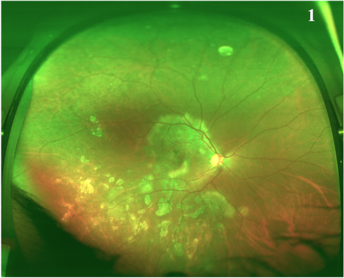
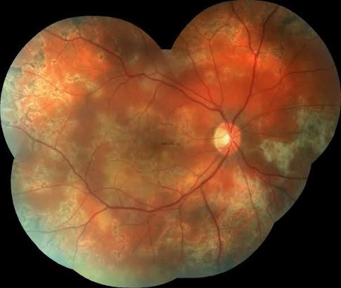
Tubercular serpiginous-like choroiditis is common in tuberculosis-endemic regions and is increasingly recognized elsewhere, where the diagnosis is easy to miss if the idiopathic serpiginous pattern is assumed without considering an infectious cause.
Tubercular Serpiginous-Like Choroiditis vs the Classic Pattern
Idiopathic serpiginous choroidopathy typically starts at the optic disc and spreads in a continuous, geographic pattern, and it is the pattern that tubercular serpiginous-like choroiditis most often mimics.
Tubercular SLC more often shows multiple discrete lesions, sometimes at the disc and sometimes away from it, and the lesions can be discontinuous instead of confluent; it may also have more vitritis than the classic form (see serpiginous choroidopathy).
Lesions can coalesce over time to resemble the classic pattern, which is part of why the two are easy to confuse without imaging and systemic workup.
Clinical and Imaging Features
- Multifocal, yellow-gray subretinal lesions, often with some vitreous haze
- Healed lesions leave chorioretinal atrophy and pigmentary change
- Fluorescein angiography shows early hypofluorescence with late staining of active lesion edges
- Indocyanine green angiography is particularly useful, showing hypofluorescent lesions that are often more extensive than what is visible clinically or on fluorescein angiography, reflecting choroidal involvement beyond the visible retinal lesion
- Retinal vasculitis and papillitis can accompany the choroiditis in some patients
Fundus Explorer Pro
Photograph the retinal findings described here with the phone already in your pocket — 22 D optics and built-in illumination in one handheld unit.
From Choroida — the team behind this siteWorkup for Tuberculosis
Given the overlapping appearance with idiopathic disease, a tuberculosis workup is warranted in any patient with a serpiginous-like choroiditis pattern, particularly from or with travel to an endemic region.
- Tuberculin skin test or interferon-gamma release assay
- Chest radiography or CT to look for evidence of pulmonary tuberculosis, present or past
- Aqueous or vitreous PCR for Mycobacterium tuberculosis in selected cases, though sensitivity is limited
- A history of tuberculosis exposure or prior treatment
A positive tuberculin or interferon-gamma result supports the diagnosis but reflects exposure rather than proof of active ocular infection, so the diagnosis remains largely clinical, supported by imaging and the systemic workup (see ocular tuberculosis).
Differential Diagnosis
- Idiopathic serpiginous choroidopathy
- Acute syphilitic posterior placoid chorioretinitis (see ASPPC)
- Relentless placoid chorioretinitis
- Multifocal choroiditis and panuveitis of other causes
- Sarcoid choroiditis
Treatment
Standard four-drug antitubercular therapy (ATT) is given for a prolonged course, typically several months to a year, often longer than for pulmonary tuberculosis alone because of the difficulty of confirming ocular cure.
Corticosteroids are usually added alongside ATT once antitubercular cover is established, since inflammation can worsen transiently as mycobacterial antigens are released, a paradoxical reaction that is managed with steroid cover rather than by stopping ATT.
Regular monitoring for medication toxicity, including liver function and visual function for ethambutol, is part of the treatment plan (see toxic and nutritional optic neuropathy for ethambutol-related optic nerve toxicity).
Prognosis
Response to ATT and corticosteroids is generally good, with inflammation quieting over weeks to months, though healed lesions leave permanent chorioretinal scarring.
Recurrence can occur, particularly if ATT is stopped early or the systemic tuberculosis diagnosis was incomplete, so close follow-up continues through and after the treatment course.


Document what you see
Two smartphone imaging tools built for everyday clinic use — one for the slit lamp, one for the fundus.
From Choroida — the team behind this siteReferences
- Gupta A, Bansal R, Gupta V, Sharma A, Bambery P. Ocular signs predictive of tubercular uveitis. Am J Ophthalmol. 2010;149:562-570.
- Gupta V, Gupta A, Rao NA. Intraocular tuberculosis: an update. Surv Ophthalmol. 2007;52:561-587.
- Bansal R, Gupta A, Gupta V, Dogra MR, Bambery P, Arora SK. Tubercular serpiginous-like choroiditis presenting as multifocal serpiginoid choroiditis. Ophthalmology. 2012;119:2334-2342.
- Agarwal A, Agrawal R, Gunasekaran DV, et al. The Collaborative Ocular Tuberculosis Study (COTS)-1 report 3: polymerase chain reaction in the diagnosis and management of tubercular uveitis. Am J Ophthalmol. 2019;198:96-105.
Test yourself
A few questions straight from this article.
-
Tubercular serpiginous-like choroiditis is best characterised as which of the following?
Tubercular serpiginous-like choroiditis is a form of ocular tuberculosis that imitates classic serpiginous choroidopathy on fundus examination but follows a different treatment path. -
Which lesion pattern favours tubercular serpiginous-like choroiditis over idiopathic serpiginous choroidopathy?
Idiopathic serpiginous choroidopathy starts at the optic disc and spreads continuously, whereas tubercular SLC shows multiple discrete lesions, sometimes away from the disc, with more vitritis. -
Which additional ocular findings can accompany tubercular serpiginous-like choroiditis?
Retinal vasculitis and papillitis can accompany the choroiditis in some patients, alongside the multifocal yellow-grey subretinal lesions and vitreous haze. -
Why is indocyanine green angiography particularly useful in tubercular serpiginous-like choroiditis?
ICG angiography shows hypofluorescent lesions often more extensive than what is visible clinically or on fluorescein angiography, reflecting choroidal involvement beyond the visible retinal lesion. -
Fluorescein angiography in tubercular serpiginous-like choroiditis shows which pattern?
Fluorescein angiography shows early hypofluorescence followed by late staining of the edges of the active lesions. -
What does a positive tuberculin skin test or interferon-gamma release assay establish in serpiginous-like choroiditis?
A positive tuberculin or interferon-gamma result supports the diagnosis but reflects exposure, so the diagnosis stays largely clinical, supported by imaging and the systemic workup. -
How long is antitubercular therapy typically given for tubercular serpiginous-like choroiditis?
Standard four-drug antitubercular therapy runs for a prolonged course of several months to a year, often longer than for pulmonary tuberculosis alone because ocular cure is hard to confirm. -
Inflammation may worsen transiently soon after antitubercular therapy begins. How is this handled?
Inflammation can flare transiently as mycobacterial antigens are released, and this paradoxical reaction is managed with corticosteroid cover rather than by stopping antitubercular therapy. -
Which monitoring belongs in the treatment plan during antitubercular therapy for ocular tuberculosis?
Regular monitoring for medication toxicity includes liver function and visual function, the latter because of ethambutol-related optic nerve toxicity. -
Which circumstance most raises the risk of recurrence in tubercular serpiginous-like choroiditis?
Recurrence is particularly likely if antitubercular therapy is stopped early or the systemic tuberculosis diagnosis was incomplete, so follow-up continues through and beyond the course.
