
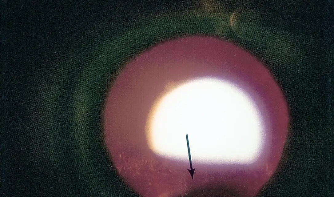
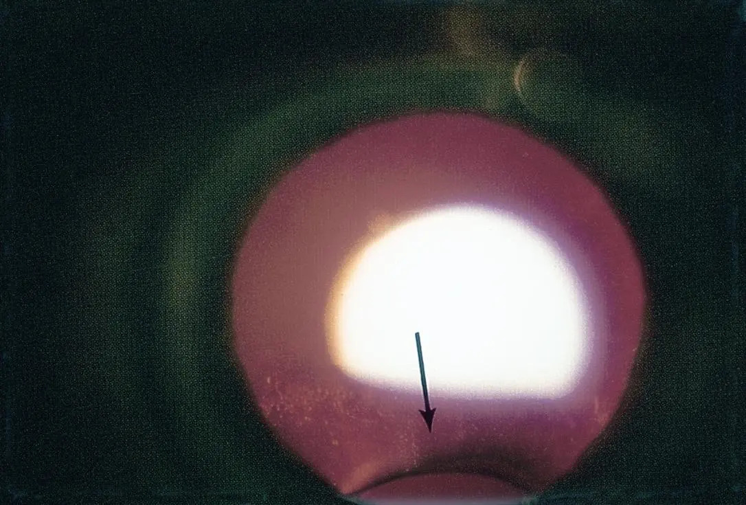
A lens coloboma is a genuinely rare finding, a notch or flattened segment at the equator of the lens, and while it shares its name and, to some extent, its embryologic story with the more familiar iris and choroidal coloboma, the mechanism producing a lens coloboma specifically involves abnormal or absent zonular fiber development in a localized area, rather than the fetal fissure closure defect responsible for coloboma elsewhere in the eye.
A Different, Zonular-Based Mechanism
While iris, choroidal, and optic disc coloboma all result from incomplete closure of the fetal fissure during early eye development, a lens coloboma arises from a more localized problem: absent or deficient zonular fibers in a specific sector, which fail to provide the normal, complete circumferential support the lens equator depends on for its round, symmetric shape.
Without this zonular support in the affected sector, the lens develops a flattened or notched equatorial contour in that specific area, distinct in mechanism from the fetal fissure defect responsible for coloboma involving the iris, choroid, and optic disc, even though these findings frequently coexist in the same patient given their common association with the broader spectrum of ocular developmental anomalies.
Clinical Appearance
- A notch or flattened area at the lens equator, best appreciated with pupillary dilation, sometimes subtle and easily overlooked unless specifically sought
- Often located inferiorly or inferonasally, though the exact position can vary
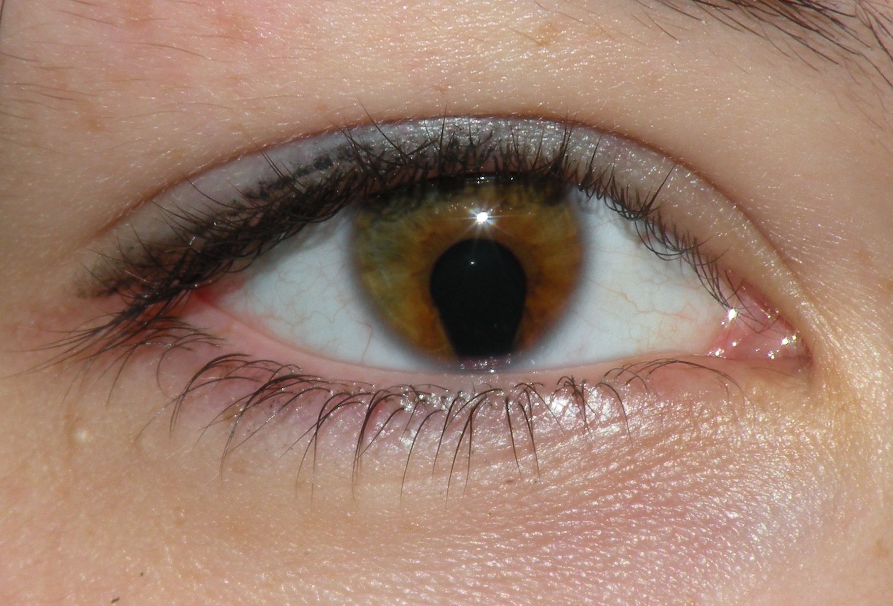
- Frequently, though not universally, associated with a coexisting iris coloboma in the same quadrant, reflecting the shared developmental field these structures arise from, even given the distinct specific mechanism producing the lens finding itself
- Can be unilateral or bilateral, and severity varies from a subtle notch to a more pronounced defect
All-fit Slit-Lamp Adapter
Record and share exactly what you see at the slit lamp. One adapter fits any slit lamp or surgical microscope — and any smartphone.
From Choroida — the team behind this siteAssociated Findings
- Iris coloboma, frequently coexisting and often in a corresponding location, given the related developmental context
- Choroidal and optic disc coloboma may also coexist, reflecting the broader spectrum of colobomatous involvement some affected eyes display
- Cataract can develop in association with a lens coloboma, sometimes related to the abnormal lens capsule and zonular support in the affected area
- As with other forms of ocular coloboma, systemic associations, including CHARGE syndrome, should be considered, particularly when lens coloboma coexists with iris or choroidal coloboma (see optic disc coloboma for a detailed discussion of the systemic associations relevant across this family of related developmental anomalies)
Clinical and Visual Significance
- A lens coloboma itself, when mild, may cause minimal visual disturbance, though larger defects can be associated with lenticular astigmatism or other refractive irregularity related to the altered lens shape
- The zonular weakness underlying a lens coloboma has direct practical relevance if cataract surgery is ever needed in that eye, since the same localized absence of zonular support that produced the coloboma also represents a zone of reduced structural support during lens extraction, requiring surgical planning adjustments similar in principle to those used for other causes of focal zonular weakness (see zonular dehiscence in cataract surgery and capsular tension ring indications for the relevant surgical planning framework)
Diagnostic Evaluation
- Careful examination with pupillary dilation to identify and characterize the lens equator notch, since this finding can be subtle and is easily missed without deliberate, complete examination of the lens periphery
- Assessment for coexisting iris, choroidal, and optic disc coloboma
- Systemic evaluation for associated syndromes, particularly when multiple ocular structures show colobomatous involvement, following the same principles applied when any form of ocular coloboma is identified
- Refraction to characterize any associated lenticular astigmatism or refractive irregularity
Management
Observation
Mild lens coloboma without significant visual impact or cataract is managed with observation, since the finding itself does not require direct treatment in the absence of a visually significant complication.
Correcting Refractive Error
Any associated refractive irregularity is corrected with spectacles or contact lenses as needed, similar to management of irregular astigmatism from other causes.
Cataract Surgery Considerations
Should cataract surgery become necessary in an eye with lens coloboma, careful preoperative recognition of the associated focal zonular weakness supports appropriate surgical planning, including consideration of capsular support devices, following the same general principles applied to other causes of zonular compromise identified before surgery.
Prognosis
Lens coloboma, particularly when mild and isolated, generally carries a good visual prognosis, with management focused on correcting any associated refractive irregularity and monitoring for cataract development over time.
As with other forms of ocular coloboma, the broader prognosis and management considerations often extend beyond the eye itself when the lens finding is part of a more extensive pattern of colobomatous involvement, reinforcing the value of a complete ocular and, when appropriate, systemic evaluation whenever this uncommon finding is identified.


Document what you see
Two smartphone imaging tools built for everyday clinic use — one for the slit lamp, one for the fundus.
From Choroida — the team behind this siteReferences
- Onwochei BC, Simon JW, Bateman JB, Couture KC, Mir E. Ocular colobomata. Surv Ophthalmol. 2000;45:175-194.
- Nelson LB, Maumenee IH. Ectopia lentis. Surv Ophthalmol. 1982;27:143-160.
- Pagon RA. Ocular coloboma. Surv Ophthalmol. 1981;25:223-236.
- Jesberg DO, Schepens CL. Retinal detachment associated with coloboma of the choroid. Arch Ophthalmol. 1961;65:163-173.
