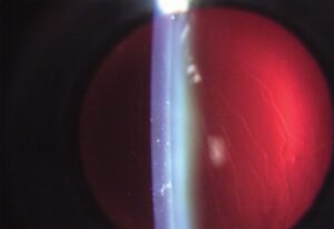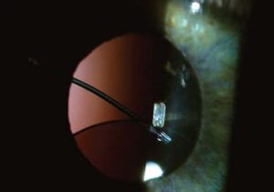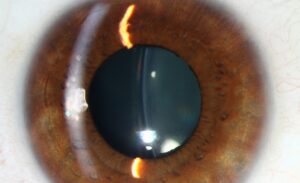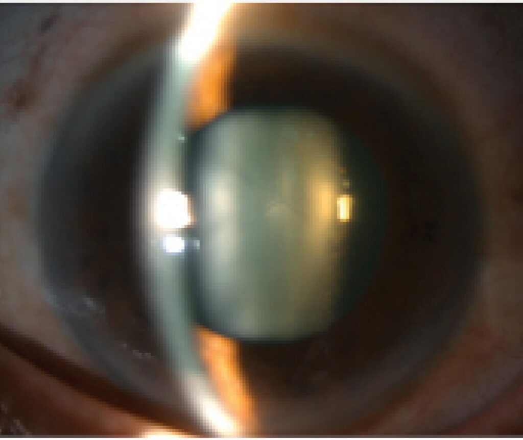
Inferonasal subluxation of lens seen in homocystinuria describes a lens that has slipped partly downward and toward the nose because its zonular fibers are weak or broken. Marfan syndrome, by contrast, classically shifts the lens superotemporally. Both patterns are usually bilateral, and recognizing the direction at the slit lamp can point you toward a systemic diagnosis the patient has not received.
What Lens Subluxation Means
Lens subluxation means partial displacement of the lens from its normal central position, caused by stretching, weakness, or focal breakdown of the zonular fibers. The lens stays at least partly within the pupillary space and often still gives useful vision. Clinicians call the congenital or inherited form ectopia lentis.
Subluxation differs from complete lens dislocation, in which zonular support has failed entirely and the lens lies in the anterior chamber or vitreous cavity. Searches for eye subluxation usually mean this condition. Dislocation is the more severe state, and it needs a different surgical plan.
Inferonasal Subluxation of Lens Seen in Homocystinuria
Homocystinuria, an autosomal recessive disorder of methionine metabolism, classically causes inferior or inferonasal lens displacement, and ectopia lentis is its most common ocular finding. The zonular fibers weaken progressively, so the lens often drifts during childhood. Patients may also have a marfanoid habitus, intellectual disability, and a tendency toward thromboembolism.
That vascular risk makes the finding matter beyond the eye. General anesthesia and dehydration can precipitate thrombosis, so tell the anesthesia team and a metabolic physician before any surgery. Dietary therapy and, in responsive patients, pyridoxine change the systemic course.
Inferior subluxation of lens also occurs in Weill-Marchesani syndrome, where small, round, microspherophakic lenses are typical, and in sulfite oxidase deficiency. In ectopia lentis et pupillae, the pupil and the lens are displaced in opposite directions.
All-fit Slit-Lamp Adapter
Record and share exactly what you see at the slit lamp. One adapter fits any slit lamp or surgical microscope — and any smartphone.
From Choroida — the team behind this siteSuperotemporal Lens Subluxation in Marfan Syndrome
Superotemporal lens subluxation is the classic pattern in Marfan syndrome, a fibrillin-1 disorder that weakens the zonular fibers. The change is usually bilateral and fairly symmetric, and the lens typically stays clear. This upward and outward direction helps separate Marfan syndrome from homocystinuria, although overlap exists and direction alone never confirms a diagnosis.
Bilateral lens subluxation without trauma should always prompt a systemic search. In suspected Marfan syndrome, arrange echocardiography for aortic root dilation and refer for genetic evaluation, because the cardiovascular risk outweighs the eye findings. Familial isolated ectopia lentis also occurs without other features.
Symptoms of a Subluxated Lens
A subluxated lens symptom picture depends on how far the lens has moved. Mild displacement may cause no complaints, while greater displacement produces blurred or fluctuating vision, and monocular diplopia when the pupil spans both the lens edge and the aphakic gap. Many patients develop lenticular myopia and irregular astigmatism.
Pain, redness, and halos suggest a complication such as angle closure or inflammation. Children rarely complain, so amblyopia, squinting, or an unusual red reflex may be the first clue, and any child with these signs deserves a dilated examination.
Lens Subluxation at the Slit Lamp
Slit lamp examination, preferably after pupillary dilation, is the main way to confirm lens subluxation. Examine both eyes, because bilateral findings change the diagnostic question entirely. Retroillumination and a careful sweep around the zonular circumference reveal most cases.
Phacodonesis
Phacodonesis is a trembling of the lens. Ask the patient to make a saccade and then stop, and watch the lens keep oscillating briefly because its zonular anchoring is inadequate. It is a direct and often the most reliable sign of zonular weakness.
Iridodonesis
Iridodonesis is a tremulous movement of the iris. The iris normally rests against the anterior lens surface, so a poorly anchored lens leaves it unsupported and it shakes with eye movement. It is also present in aphakic eyes.
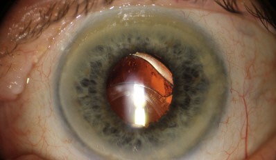
Asymmetric Anterior Chamber Depth
A tilted or displaced lens can shallow or deepen the anterior chamber depending on the direction of displacement, and depth may differ between clock hours in the same eye. Compare the two eyes, and a very shallow chamber raises concern for pupillary block.
Visible Lens Edge
With greater displacement, the lens equator and sometimes stretched or broken zonular fibers appear within the dilated pupil. Seeing the edge confirms the diagnosis directly. Vitreous may prolapse forward through the gap, and the aphakic and phakic parts of the pupil focus light differently.
Causes of Unilateral and Bilateral Lens Subluxation
Bilateral subluxation usually points to a genetic or metabolic cause. Unilateral subluxation more often follows trauma or local disease, so the history and the pattern together narrow the list quickly. The common causes are listed below.
- Marfan syndrome: bilateral, usually superotemporal displacement from a fibrillin-1 defect.
- Homocystinuria: bilateral, usually inferior or inferonasal displacement.
- Weill-Marchesani syndrome: small spherical lens, often displaced inferiorly, with a risk of pupillary block.
- Trauma: zonular dialysis at the injury site, so the lens shifts toward the intact zonules and away from the tear, usually in one eye.
- Pseudoexfoliation syndrome: gradual, often asymmetric zonular weakness in older adults, with high intraoperative risk.
- Other causes: Ehlers-Danlos syndrome, sulfite oxidase deficiency, ectopia lentis et pupillae, aniridia, high myopia, hypermature cataract, and intraocular tumors.
Diagnostic Workup
Start with a directed history covering trauma, prior eye surgery, family history, and systemic features such as tall stature, joint laxity, or thrombotic events. Then confirm the diagnosis at the slit lamp and look for complications before planning treatment.
- Measure intraocular pressure and perform gonioscopy to look for angle closure or angle recession.
- Perform a dilated fundus examination for retinal tears or detachment, which occur more often in these eyes.
- Use ultrasound biomicroscopy or anterior segment OCT to define the extent of zonular loss.
- Refract carefully, since lens-induced myopia and astigmatism are common.
- In bilateral cases, request echocardiography, plasma total homocysteine, and genetic testing as directed.
Complications and Surgical Implications
Subluxated lenses can cause pupillary block glaucoma and retinal detachment. Lens material released through a damaged capsule can trigger lens-induced uveitis. After blunt injury, also look for hyphema and angle recession, and treat these problems before elective surgery.
Recognize subluxation before cataract surgery, because it changes the surgical plan. Capsular tension rings, capsular hooks, modified technique, and alternative lens fixation, such as scleral or iris fixation, may be needed when zonular loss is large. A related problem is covered in our case study of a dislocated intraocular lens (IOL).
Missing the diagnosis and using standard technique raises the risk of posterior capsule rupture, vitreous loss, and nuclear fragment dislocation. Preoperative slit lamp examination, plus a surgeon prepared with capsular support devices, reduces that risk.
Prognosis
With correct preoperative recognition and appropriate surgical planning, good visual outcomes are achievable even in eyes with significant zonular compromise. Spectacles or contact lenses may suffice for mild subluxation, and patients with stable, mild displacement may need only observation.
Beyond the eye, identifying bilateral subluxation, especially with a Marfan or homocystinuria pattern, carries broad diagnostic value. These conditions carry serious cardiovascular and thrombotic risks, so prompt referral can protect the patient’s life as well as vision.


Document what you see
Two smartphone imaging tools built for everyday clinic use — one for the slit lamp, one for the fundus.
From Choroida — the team behind this siteReferences
- Nelson LB, Maumenee IH. Ectopia lentis. Surv Ophthalmol. 1982;27:143-160.
- Cross HE, Jensen AD. Ocular manifestations in the Marfan syndrome and homocystinuria. Am J Ophthalmol. 1973;75:405-420.
- Jarrett WH 2nd. Dislocation of the lens: a study of 166 hospitalized cases. Arch Ophthalmol. 1967;78:289-296.
- Halpert M, BenEzra D. Surgery of the hereditary subluxated lens in children. Ophthalmology. 1996;103:681-686.
Test yourself
A few questions straight from this article.
-
Lens subluxation is best defined as which of the following?
Subluxation is partial displacement caused by weakness or focal breakdown of the zonular fibres. In complete dislocation the zonular support has failed entirely and the lens leaves the pupillary space. -
Which slit-lamp finding is the most reliable direct sign of zonular weakness in lens subluxation?
Phacodonesis — the lens continuing to wobble once the eye has stopped moving — reflects inadequate zonular anchoring and is the most reliable sign of zonular weakness. -
Why does iridodonesis develop in eyes with lens subluxation?
The iris normally rests against and is partly supported by the anterior lens surface, so a poorly anchored lens allows the iris to tremble more than normal with eye movement. -
Marfan syndrome classically produces lens subluxation in which direction?
Marfan syndrome arises from a fibrillin connective tissue abnormality affecting the zonules and classically gives bilateral, often symmetric superotemporal subluxation. -
Which direction of lens subluxation points to homocystinuria rather than Marfan syndrome?
Homocystinuria classically causes inferonasal subluxation, the opposite direction to the superotemporal pattern of Marfan syndrome, and this helps separate the two clinically. -
In traumatic lens subluxation, the lens typically displaces in which relation to the impact?
Trauma tears the zonules focally at the site of impact, producing asymmetric, often unilateral subluxation directed away from the traumatic force. -
Which condition commonly causes generalized, gradual zonular weakness relevant to cataract surgery planning?
Pseudoexfoliation syndrome produces more generalized, gradual zonular weakness and carries a recognised intraoperative risk, so it matters when planning cataract surgery. -
Which investigation best characterizes the extent and location of zonular loss before surgery?
Gonioscopy and, when needed, ultrasound biomicroscopy define how much zonular support has been lost and where, which is what surgical planning depends on. -
Which intraoperative complications rise when lens subluxation is missed before standard cataract surgery?
Proceeding with standard technique in an unrecognised subluxated lens significantly increases the risk of posterior capsular rupture and dislocation of lens fragments. -
Recognizing lens subluxation before cataract surgery should prompt consideration of which measure?
Preoperative recognition changes the surgical approach, prompting capsular support devices, modified technique and, in severe cases, alternative intraocular lens fixation.

Writes and reviews the clinical articles on Educate. Every post is checked against peer-reviewed sources before it goes live.