
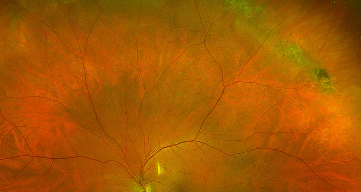
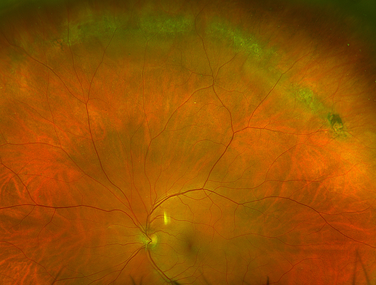
Lattice degeneration with holes describes a specific and common variant of lattice degeneration in which one or more round, atrophic breaks have already formed within the thinned lattice lesion itself, and understanding why these atrophic holes generally behave less aggressively than a traction-related flap tear is central to making sound, evidence-based decisions about whether prophylactic treatment is actually needed.
Lattice Degeneration as a Starting Point
Lattice degeneration is a common peripheral retinal finding characterized by an area of retinal thinning, often oval or linear in shape, with a criss-crossing pattern of sclerosed retinal vessels within the lesion (giving the condition its name) and firm, abnormal vitreoretinal adhesion at the margins of the lesion, a combination that makes lattice degeneration a recognized risk factor for retinal tear formation, particularly at the time of an acute posterior vitreous detachment.
How Atrophic Holes Differ From Flap Tears
Within an area of lattice degeneration, the chronically thinned retina can spontaneously develop a round, atrophic hole, essentially forming from gradual tissue thinning and breakdown rather than from an acute traction event, and this developmental mechanism is precisely why atrophic holes within lattice degeneration are generally considered to carry a lower risk of progressing to clinically significant retinal detachment than a flap (horseshoe) tear, which results from acute vitreous traction actively pulling the retina open at the time of a posterior vitreous detachment.
Fundus Explorer Pro
Photograph the retinal findings described here with the phone already in your pocket — 22 D optics and built-in illumination in one handheld unit.
From Choroida — the team behind this siteRisk Stratification
- Atrophic holes within lattice degeneration, without any associated subretinal fluid and without acute symptoms, are generally considered lower risk and are frequently managed with observation alone in an asymptomatic patient
- The presence of any surrounding or associated subretinal fluid extending from an atrophic hole changes the risk calculation substantially and generally warrants treatment, similar to the approach for a symptomatic flap tear
- Additional risk factors that shift the balance toward treatment even for an otherwise lower-risk atrophic hole include high myopia, a history of retinal detachment in the fellow eye, a strong family history of retinal detachment, and any planned intraocular surgery, particularly cataract surgery, which can itself alter vitreous dynamics and increase subsequent detachment risk
Clinical Evaluation
- Dilated fundus examination with scleral depression to fully characterize the extent of lattice degeneration, the number and size of any associated atrophic holes, and the presence or absence of any surrounding subretinal fluid
- Assessment of symptoms, since new, acute flashes and floaters in a patient with known lattice degeneration and atrophic holes should prompt particularly careful reevaluation, given the possibility of a new, acute event superimposed on the pre-existing chronic finding
- Consideration of the broader risk factor profile, including refractive error, fellow eye history, and any planned intraocular surgery, when weighing observation against prophylactic treatment for an asymptomatic finding
Management
Observation
Asymptomatic atrophic holes within lattice degeneration, without associated subretinal fluid and without other significant additional risk factors, are frequently managed with observation and routine periodic monitoring, reflecting the generally more indolent natural history of this specific pattern compared with an acute flap tear.
Prophylactic Treatment
When treatment is chosen, whether due to associated subretinal fluid, symptomatic presentation, or a combination of additional risk factors favoring a more cautious approach, laser retinopexy or cryotherapy is applied around the lesion to create a chorioretinal adhesion, aiming to wall off the area and prevent any future progression to clinically significant retinal detachment.
Individualized Decision-Making
The decision between observation and prophylactic treatment for an asymptomatic atrophic hole within lattice degeneration is individualized, weighing the generally lower inherent risk of this specific finding against the patient’s broader risk factor profile and personal preferences, rather than following a single uniform rule applied to every patient with this finding.
Patient Education
Regardless of the specific management decision for an existing lattice degeneration and atrophic hole finding, education about the symptoms of an acute posterior vitreous detachment or retinal detachment, new flashes, a sudden increase in floaters, or a curtain-like visual field defect, remains an essential part of counseling, since these symptoms would warrant urgent reevaluation regardless of the baseline management approach chosen.
Prognosis
The majority of patients with asymptomatic atrophic holes within lattice degeneration, managed with observation, never progress to clinically significant retinal detachment, supporting the generally conservative approach taken for this specific, lower-risk pattern.
For the smaller number of patients who do require treatment, whether due to associated fluid, symptoms, or additional risk factors, prophylactic laser or cryotherapy is generally effective at preventing progression to detachment, provided it is applied before significant subretinal fluid has already accumulated and spread.


Document what you see
Two smartphone imaging tools built for everyday clinic use — one for the slit lamp, one for the fundus.
From Choroida — the team behind this siteReferences
- Byer NE. Long-term natural history of lattice degeneration of the retina. Ophthalmology. 1989;96:1396-1401.
- Wilkinson CP. Evidence-based analysis of prophylactic treatment of asymptomatic retinal breaks and lattice degeneration. Ophthalmology. 2000;107:12-15.
- Folk JC, Arrindell EL, Klugman MR. The fellow eye of patients with phakic lattice retinal detachment. Ophthalmology. 1989;96:72-79.
- Byer NE. What happens to untreated asymptomatic retinal breaks, and are they affected by posterior vitreous detachment? Ophthalmology. 1998;105:1045-1049.
