
Cavernous sinus syndrome describes the combination of findings that results from a lesion within the cavernous sinus, where cranial nerves III, IV, V1, V2, and VI, along with the internal carotid artery and its sympathetic plexus, all pass through a confined space.
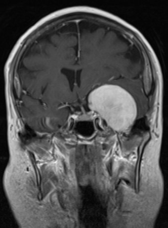
Because so many structures are packed together in one location, a single lesion here tends to produce a recognizable multi-nerve pattern rather than an isolated palsy, and recognizing that pattern is often the fastest way to localize the problem before imaging is even reviewed.
Anatomy That Explains the Pattern
The cavernous sinus sits on either side of the pituitary gland and sella turcica, and within its walls or lumen run the oculomotor (III), trochlear (IV), ophthalmic and maxillary divisions of the trigeminal (V1, V2), and abducens (VI) nerves, along with the internal carotid artery and sympathetic fibers.
A lesion of sufficient size anywhere in this space can affect any combination of these structures, which is why cavernous sinus syndrome so often presents with more than one cranial nerve involved at once, along with sometimes a Horner syndrome from disruption of the sympathetic fibers.
Clinical Features
- Diplopia from combined third, fourth, and sixth nerve involvement, which can produce a nearly immobile eye in severe cases
- Ptosis and a dilated, poorly reactive pupil if the third nerve’s pupillary fibers are affected
- Facial pain or numbness in the V1 or V2 distribution
- Proptosis if there is associated venous congestion or an expanding mass
- Horner syndrome, from disruption of sympathetic fibers traveling with the carotid artery, which can coexist confusingly with a third nerve palsy if both are affected
The combination of ophthalmoplegia with facial sensory loss and orbital signs, rather than an isolated cranial nerve palsy, is the clinical hallmark that should prompt imaging of the cavernous sinus and skull base specifically.
Causes
- Pituitary apoplexy, hemorrhage or infarction of a pituitary adenoma expanding laterally into the cavernous sinus (see pituitary apoplexy)
- Cavernous sinus thrombosis, often from spreading infection of the face or sinuses, a true emergency (see cavernous sinus thrombosis)
- Carotid-cavernous fistula, producing a pulsatile, congested pattern with a bruit (see carotid-cavernous fistula)
- Tolosa-Hunt syndrome, an idiopathic granulomatous inflammation of the cavernous sinus, a diagnosis of exclusion
- Tumors, including meningioma, nasopharyngeal carcinoma with skull base extension, schwannoma, and metastatic disease
- Aneurysm of the intracavernous carotid artery
Workup
- MRI of the brain and orbits with contrast, focused on the cavernous sinus and skull base, is the primary imaging study
- MR or CT angiography when a vascular cause, such as aneurysm or fistula, is suspected
- Endocrine evaluation when pituitary disease is suspected
- Laboratory and inflammatory markers, and sometimes biopsy, when Tolosa-Hunt syndrome or a systemic inflammatory process is considered, since it remains a diagnosis of exclusion
Urgency depends on the suspected cause: cavernous sinus thrombosis and pituitary apoplexy with visual compromise need immediate evaluation and treatment, while a slowly progressive tumor allows a more measured workup.
Management
Treatment follows the underlying cause: antibiotics and anticoagulation for septic thrombosis, urgent endocrine and sometimes surgical management for pituitary apoplexy, endovascular treatment for a fistula or aneurysm, corticosteroids for Tolosa-Hunt syndrome after malignancy and infection have been reasonably excluded, and surgery or radiotherapy for tumors.
Ocular management includes lubrication and, for persistent diplopia, prism correction or strabismus surgery once the underlying condition is controlled and stable.
Prognosis
Outcome depends entirely on the cause and how quickly it is treated, ranging from full recovery after resolution of an inflammatory or infectious process to permanent cranial nerve deficits from tumor invasion or a severe vascular event.
The multi-nerve pattern of cavernous sinus syndrome is valuable precisely because it narrows the differential quickly, directing the workup toward a small set of possible causes rather than a broad neurological survey.


Document what you see
Two smartphone imaging tools built for everyday clinic use — one for the slit lamp, one for the fundus.
From Choroida — the team behind this siteReferences
- Keane JR. Cavernous sinus syndrome: analysis of 151 cases. Arch Neurol. 1996;53:967-971.
- Fernandez S, Godino O, Martinez-Yelamos S, et al. Cavernous sinus syndrome: a series of 126 patients. Medicine (Baltimore). 2007;86:278-281.
- Kline LB, Hoyt WF. The Tolosa-Hunt syndrome. J Neurol Neurosurg Psychiatry. 2001;71:577-582.
- Bhatti MT. Cavernous sinus syndrome. In: Neuro-Ophthalmology Review Manual. 7th ed. Thorofare, NJ: Slack; 2013.
