
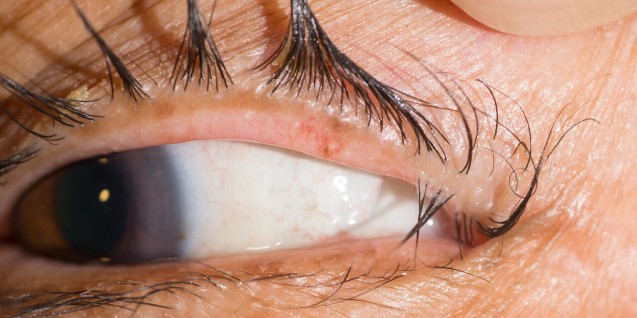
Trichiasis is misdirection of otherwise normally positioned eyelashes so that they rub against the ocular surface, causing chronic irritation and, if untreated long enough, real corneal damage.
It is distinct from entropion, where the entire eyelid margin turns inward and carries otherwise normally oriented lashes with it, even though the two conditions can look similar at a glance and sometimes coexist in the same patient.
That distinction matters directly for treatment, since fixing an inward-turned lid margin does nothing for lashes that are simply growing in the wrong direction from an otherwise normally positioned lid.
Trichiasis is common enough worldwide that it represents a genuine public health problem in regions with endemic trachoma, where it is a leading preventable cause of corneal blindness.
Mechanism
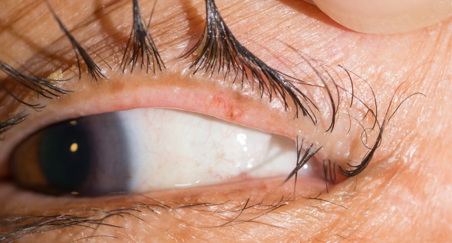
In trichiasis, the eyelid margin itself sits in its normal anatomic position, but individual lashes emerge at an abnormal angle, typically inward toward the globe, so that the lash tips contact the cornea or conjunctiva with each blink.
This is fundamentally different from entropion, where the lash misdirection is a secondary consequence of the entire lid margin rotating inward, or from distichiasis, where an entire extra row of lashes emerges from the meibomian gland orifices, both discussed in their own dedicated articles on this site.
Chronic mechanical rubbing from misdirected lashes causes corneal epithelial irritation that, over time, can progress to punctate keratopathy, corneal scarring, or, in severe or longstanding cases, neovascularization.
The number of affected lashes can range from a single stray lash to dozens across the lid margin, and symptom severity does not always track cleanly with how many lashes are actually involved.
Causes
- Chronic blepharitis and lid margin inflammation, the most common cause in general practice, disrupting normal lash follicle orientation over time
- Trachoma, a leading cause worldwide in endemic regions, where recurrent conjunctival scarring distorts the lid margin and follicle architecture
- Herpes zoster ophthalmicus or other cicatrizing conjunctival disease, producing localized scarring that redirects affected lashes
- Chronic mechanical or thermal injury to the lid margin, including prior eyelid surgery or trauma
- Idiopathic trichiasis, where no clear underlying cause is identified despite a thorough history and exam
All-fit Slit-Lamp Adapter
Record and share exactly what you see at the slit lamp. One adapter fits any slit lamp or surgical microscope — and any smartphone.
From Choroida — the team behind this siteClinical Presentation
Patients typically report foreign body sensation, tearing, and photophobia, symptoms that can be difficult to distinguish from dry eye disease or blepharitis without a careful lid margin exam specifically looking for misdirected lashes.
Corneal findings range from mild superficial punctate staining in early or mild cases to frank corneal ulceration, scarring, or neovascularization in longstanding, untreated trichiasis, particularly when lashes contact the visual axis.
A careful slit-lamp exam with lid eversion and magnification is needed to identify individual misdirected lashes, since a small number of abnormal lashes can cause disproportionate symptoms and corneal damage relative to how subtle they may look on cursory exam.
Fluorescein staining helps localize exactly where a misdirected lash is contacting the ocular surface, which is especially useful when only one or two lashes among many are actually responsible for the patient’s symptoms.
Management
Simple epilation, manually removing the offending lashes with forceps, provides immediate relief but is temporary, since lashes typically regrow within four to six weeks in their original, misdirected orientation.
Electrolysis or thermal ablation of the follicle offers more durable removal for a small number of isolated misdirected lashes, destroying the follicle itself rather than just the visible lash.
Cryotherapy applied to the lid margin can treat a broader area of trichiasis at once, though it carries some risk of depigmentation and, if overly aggressive, damage to adjacent normal lashes and meibomian gland function.
Surgical options, including lid margin rotation procedures, are reserved for extensive or recurrent trichiasis, particularly when an underlying cicatricial process is actively distorting the lid margin architecture and simpler measures keep failing.
Any underlying cause, such as active blepharitis or trachoma, needs its own targeted treatment alongside management of the trichiasis itself, since treating the lashes without addressing an ongoing inflammatory or infectious driver invites recurrence.
Patients with recurrent trichiasis benefit from understanding that some degree of ongoing surveillance is often needed even after successful treatment, since new lashes can become misdirected over time even when the original offending lashes are permanently resolved.


Document what you see
Two smartphone imaging tools built for everyday clinic use — one for the slit lamp, one for the fundus.
From Choroida — the team behind this siteReferences
- American Academy of Ophthalmology. Basic and Clinical Science Course, Section 7: Oculofacial Plastic and Orbital Surgery.
- Bowling B. Kanski’s Clinical Ophthalmology.
- Hardten DR, Meyer DR, Yeatts RP. Trichiasis: pathogenesis and treatment options. Ophthalmology Clinics of North America.
Test yourself
A few questions straight from this article.
-
Trichiasis is best defined as which eyelid abnormality?
In trichiasis the lid margin sits in its normal anatomic position while individual lashes emerge at an abnormal angle and rub the ocular surface with each blink. -
In entropion, what causes the eyelashes to contact the globe?
Entropion turns the entire lid margin inward while the lashes themselves remain normally oriented. That is why correcting margin position treats entropion but does nothing for true trichiasis. -
In which setting is trichiasis a leading preventable cause of corneal blindness?
Trichiasis is a genuine public health problem in areas with endemic trachoma, where recurrent conjunctival scarring distorts the lid margin and follicle architecture. -
In distichiasis, where does the abnormal extra row of eyelashes emerge?
Distichiasis is defined by an entire accessory row of lashes arising from the meibomian gland orifices, which separates it from trichiasis and from entropion. -
Which condition is the most common cause of trichiasis in general ophthalmic practice?
Chronic blepharitis and lid margin inflammation disrupt normal lash follicle orientation over time, making it the commonest driver of trichiasis outside trachoma-endemic regions. -
After simple epilation for trichiasis, misdirected lashes typically regrow within what interval?
Forceps epilation relieves symptoms immediately but is only temporary, as lashes regrow in their original misdirected orientation within about four to six weeks. -
Which treatment gives durable removal of a few isolated misdirected lashes in trichiasis?
Electrolysis or thermal ablation destroys the follicle itself rather than just the visible lash, making it suited to a small number of isolated misdirected lashes. -
Cryotherapy of the lid margin for trichiasis carries which recognised risk?
Cryotherapy treats a broader area at once but risks depigmentation and, if applied too aggressively, damage to adjacent normal lashes and to meibomian gland function. -
Which test localises exactly where a misdirected lash contacts the ocular surface?
Fluorescein pinpoints the site of contact, which is especially useful when only one or two lashes among many are responsible for the patient's symptoms. -
Longstanding untreated trichiasis can progress to which corneal complication?
Chronic mechanical rubbing causes epithelial irritation that can advance from punctate keratopathy to corneal scarring and, in severe or longstanding cases, neovascularization.
