Elevated episcleral venous pressure is a less common but important mechanism of secondary glaucoma, and it illustrates a broader principle worth understanding: because aqueous humor ultimately drains into the episcleral veins, any process that raises pressure downstream in this venous system, even from a cause located well outside the eye itself, transmits backward and raises intraocular pressure, regardless of how normally the trabecular meshwork itself is functioning.
Why Episcleral Venous Pressure Matters for IOP
Aqueous humor exits the eye through the trabecular meshwork into Schlemm’s canal and then into the episcleral venous system, which itself drains into the orbital and, ultimately, systemic venous circulation.
Normal intraocular pressure depends on there being an adequate pressure gradient favoring outflow from the eye into this venous system, and when episcleral venous pressure itself is abnormally elevated, this gradient is reduced or reversed, directly raising intraocular pressure even when the trabecular meshwork’s own outflow function is entirely normal.
Causes
- Carotid-cavernous fistula, an abnormal connection between the carotid artery and cavernous sinus, causing markedly elevated pressure in the orbital venous system that drains into it, and one of the more dramatic and recognizable causes given its associated pulsatile proptosis and other orbital findings
- Sturge-Weber syndrome, a neurocutaneous syndrome with abnormal episcleral and choroidal vascular malformations, commonly associated with glaucoma related in part to elevated episcleral venous pressure from these vascular anomalies
- Superior vena cava obstruction and other causes of systemic venous congestion affecting the head and neck
- Orbital venous malformations and varices
- Thyroid eye disease, in which orbital congestion can secondarily elevate episcleral venous pressure
- Idiopathic elevated episcleral venous pressure, a rare condition in which no specific underlying cause is identified despite thorough evaluation
All-fit Slit-Lamp Adapter
Record and share exactly what you see at the slit lamp. One adapter fits any slit lamp or surgical microscope — and any smartphone.
From Choroida — the team behind this siteClinical Features
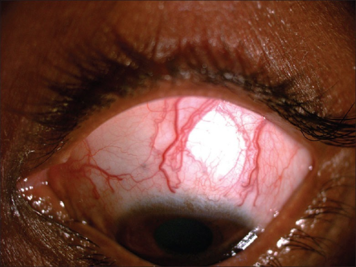
- Dilated, tortuous episcleral vessels, often a striking and clinically important examination finding, sometimes described as having a distinctive corkscrew appearance
- Elevated intraocular pressure, which can be significant, along with a relatively open angle on gonioscopy, since the mechanism is downstream of the trabecular meshwork rather than a primary angle abnormality
- Findings related to the underlying cause, such as pulsatile proptosis and an audible bruit in carotid-cavernous fistula, or a facial port-wine stain and other neurocutaneous findings in Sturge-Weber syndrome
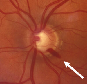
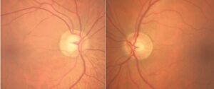
- Optic nerve damage and visual field loss can develop over time, similar to other forms of chronically elevated intraocular pressure, if the condition is not adequately recognized and managed
Diagnosis
- Careful slit-lamp examination specifically noting the appearance of episcleral vessels, since dilated, tortuous episcleral veins are a key clue pointing toward this mechanism rather than a primary trabecular meshwork problem
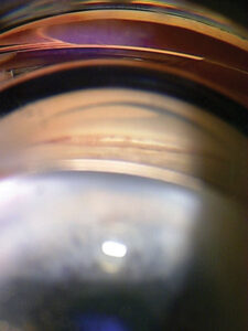
- Gonioscopy, typically showing an open, normal-appearing angle, supporting a downstream, rather than angle-based, mechanism
- Evaluation for an underlying systemic or orbital cause, including orbital and cerebrovascular imaging when carotid-cavernous fistula is suspected, and broader systemic evaluation when Sturge-Weber syndrome or another underlying condition is considered
- Direct measurement of episcleral venous pressure is possible with specialized instrumentation but is not routinely performed in most clinical settings, with the diagnosis more often made based on the characteristic clinical picture
Management
Treating the Underlying Cause
When a specific, treatable underlying cause is identified, such as a carotid-cavernous fistula, definitive treatment of that condition, often through interventional or neurosurgical closure of the fistula, can directly reduce episcleral venous pressure and improve the associated glaucoma.
Medical IOP-Lowering Therapy
Standard aqueous-suppressant glaucoma medications are used to help control intraocular pressure while the underlying cause is being addressed or when a definitive treatable cause is not identified, though the elevated episcleral venous pressure itself can limit the effectiveness of outflow-based glaucoma medications given the ongoing pressure gradient problem.
Surgical Considerations
Glaucoma surgery, including trabeculectomy, can be complicated in this setting by an increased risk of intraoperative and postoperative choroidal effusion or hemorrhage, related to the same elevated venous pressure affecting the eye more broadly, so these cases require careful surgical planning and, when possible, are approached after addressing the underlying venous pressure elevation when feasible.
Prognosis
Outcomes depend heavily on whether the underlying cause of elevated episcleral venous pressure can be identified and effectively treated, with cases related to a correctable vascular abnormality, such as a carotid-cavernous fistula, generally having a better prognosis for glaucoma control once the underlying lesion is addressed.
Chronic, ongoing causes, such as Sturge-Weber syndrome, often require sustained, sometimes lifelong glaucoma management, with attention to the technical challenges elevated episcleral venous pressure poses for both medical and surgical glaucoma treatment.


Document what you see
Two smartphone imaging tools built for everyday clinic use — one for the slit lamp, one for the fundus.
From Choroida — the team behind this siteReferences
- Minckler DS, Shammas A, Wilcox M, Ogden TE. Experimental glaucoma. Arch Ophthalmol. 1978;96:1274-1285.
- Talusan ED, Schwartz B. Episcleral venous pressure. Preliminary report on association with age and primary open-angle glaucoma. Arch Ophthalmol. 1981;99:824-828.
- Sanke RF. Increased episcleral venous pressure and glaucoma secondary to Sturge-Weber syndrome. J Pediatr Ophthalmol Strabismus. 1984;21:243-245.
- Phelps CD, Thompson HS, Ossoinig KC. The diagnosis and prognosis of atypical carotid-cavernous fistula (red-eyed shunt syndrome). Am J Ophthalmol. 1982;93:423-436.
Test yourself
A few questions straight from this article.
-
Why does elevated episcleral venous pressure raise intraocular pressure?
Normal pressure depends on a gradient favouring outflow into the episcleral veins; when venous pressure rises, that gradient falls or reverses and intraocular pressure climbs even with a perfectly functioning meshwork. -
Through which route does aqueous humour reach the episcleral venous system?
Aqueous exits via the trabecular meshwork into Schlemm's canal and then the episcleral venous system, which drains into the orbital and ultimately systemic venous circulation. -
What does gonioscopy typically show in elevated episcleral venous pressure glaucoma?
Because the obstruction lies downstream of the meshwork rather than in the angle itself, gonioscopy usually shows an open, normal-looking angle despite significantly raised pressure. -
Which slit-lamp finding points to an episcleral venous cause of raised intraocular pressure?
Dilated, tortuous episcleral veins, sometimes described as corkscrew, are the key clue steering the clinician toward a downstream venous mechanism rather than a primary meshwork problem. -
Which cause of raised episcleral venous pressure presents with pulsatile proptosis and an audible bruit?
A carotid-cavernous fistula, an abnormal connection between carotid artery and cavernous sinus, raises orbital venous pressure markedly and is recognisable by pulsatile proptosis and a bruit. -
Which vascular abnormality links Sturge-Weber syndrome to raised episcleral venous pressure?
Sturge-Weber syndrome features abnormal episcleral and choroidal vascular malformations, and the glaucoma it commonly causes relates in part to the resulting elevated episcleral venous pressure. -
Why is episcleral venous pressure rarely measured directly in clinical practice?
Direct measurement is possible with specialised instrumentation but is not routinely performed in most settings, so the diagnosis usually rests on the characteristic clinical picture. -
Why may outflow-based glaucoma medications underperform when episcleral venous pressure is raised?
The elevated venous pressure keeps the outflow gradient problem in place, which limits what outflow-based medications can achieve; aqueous suppressants are used to control pressure meanwhile. -
Which complication risk is increased when performing trabeculectomy in these eyes?
The same elevated venous pressure raises the risk of intraoperative and postoperative choroidal effusion or haemorrhage, so these cases need careful planning and, where feasible, treatment of the venous pressure first. -
Which cause of episcleral venous pressure glaucoma carries the better prognosis for pressure control?
Outcomes hinge on whether the cause is correctable: a fistula amenable to closure generally has a better prognosis for glaucoma control, while chronic causes such as Sturge-Weber often need lifelong management.

Writes and reviews the clinical articles on Educate. Every post is checked against peer-reviewed sources before it goes live.