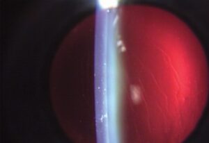
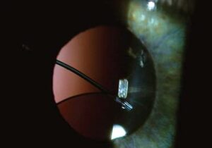
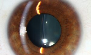
Cataract morphology, the specific location and pattern of lens opacification, is far more than a descriptive detail, since where a cataract forms shapes the symptoms a patient reports, correlates with specific underlying causes and risk factors, and can even predict how quickly it is likely to progress, all of which is why slit-lamp classification into recognized morphologic types remains central to cataract evaluation.
Nuclear Sclerotic Cataract
The most common pattern associated with normal aging, nuclear sclerotic cataract involves progressive yellowing and hardening of the central lens nucleus.
- Progresses slowly over years, typically producing a gradual, symmetric decline in vision
- Often causes a myopic shift in refraction as the nucleus increases in refractive index, sometimes producing the phenomenon of “second sight,” a temporary subjective improvement in near vision as the eye becomes more myopic
- Color vision and contrast sensitivity can be affected before visual acuity itself declines substantially, given the yellowing filter effect of the nuclear change
Cortical Cataract
This pattern involves opacification of the lens cortex, the outer layers surrounding the nucleus, classically producing wedge-shaped or spoke-like opacities extending from the lens periphery toward the center.
- Symptoms can include glare and disability under bright light conditions, sometimes disproportionate to the degree of central visual acuity loss, particularly when spokes extend into the visual axis
- Progression can be variable and sometimes asymmetric between the two eyes
- Associated with diabetes as a specific risk factor, among other general age-related risk factors
All-fit Slit-Lamp Adapter
Record and share exactly what you see at the slit lamp. One adapter fits any slit lamp or surgical microscope — and any smartphone.
From Choroida — the team behind this sitePosterior Subcapsular Cataract
Located just anterior to the posterior lens capsule, this pattern has a disproportionate functional impact relative to its often modest size, given its position directly in the path of light converging toward the retina.
- Causes prominent glare and difficulty in bright light or with pupillary constriction (such as when reading or in bright sunlight), since the central posterior location is maximally in the visual axis exactly when the pupil is smallest
- Near vision is often more affected than distance vision, related to the same pupillary constriction effect during near work
- Tends to progress more rapidly than nuclear or cortical cataract in many cases
- Associated with specific risk factors beyond simple aging, including chronic corticosteroid use (topical, systemic, or inhaled), prior ocular trauma, uveitis, and radiation exposure, making its presence, particularly in a younger patient, a prompt to specifically ask about these exposures
Congenital and Other Morphologic Patterns
- Sutural cataracts, following the Y-shaped suture lines of the lens, and other congenital morphologic patterns have their own specific associations and implications, generally evaluated in the context of pediatric cataract assessment (see pediatric cataract surgery)
- Christmas tree cataract, with colorful, polychromatic crystalline deposits, is a recognized pattern sometimes associated with myotonic dystrophy
- Mixed morphology, combining features of more than one pattern, is common, particularly in older patients where age-related nuclear and cortical change frequently coexist
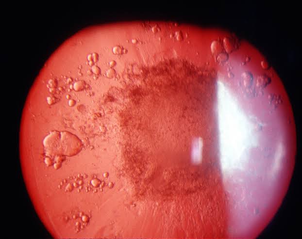
Why Morphology Guides Clinical Reasoning
- A posterior subcapsular pattern in a younger patient should prompt specific questions about corticosteroid use, prior trauma, uveitis, or radiation exposure, since this pattern is disproportionately associated with these secondary causes compared with simple age-related change
- The specific symptom pattern reported, whether predominantly glare, reduced near vision, or gradual overall blur, can be anticipated based on the morphologic type observed, helping to confirm that a patient’s symptoms match the examination findings
- Anticipated progression rate, informed by morphologic type, supports realistic counseling about the expected timeline to visually significant cataract and the point at which surgery is likely to become appropriate
Examination Technique
Careful slit-lamp examination with a dilated pupil, using both diffuse and retroillumination techniques, allows accurate classification of the specific morphologic pattern or patterns present, informing both the clinical reasoning above and objective documentation for tracking progression over subsequent visits.
Clinical Significance
Recording cataract morphology specifically, rather than simply noting “cataract present,” supports more precise counseling about expected symptoms and progression, prompts appropriate additional history-taking when a pattern suggests a secondary cause, and provides an objective baseline for monitoring change over time.
Related Reading
- Christmas Tree Cataract
- Snowflake Cataract
- Cerulean Cataract
- Rosette-Shaped Cataract
- Posterior Subcapsular Cataract
- Cortical Cataract
- Morgagnian Cataract
- Intumescent Cataract


Document what you see
Two smartphone imaging tools built for everyday clinic use — one for the slit lamp, one for the fundus.
From Choroida — the team behind this siteReferences
- Chylack LT Jr, Wolfe JK, Singer DM, et al. The Lens Opacities Classification System III. Arch Ophthalmol. 1993;111:831-836.
- Klein BE, Klein R, Linton KL. Prevalence of age-related lens opacities in a population. Ophthalmology. 1992;99:546-552.
- Harding JJ, Harding RS, Egerton M. Risk factors for cataract in Oxfordshire: diabetes, peripheral neuropathy, myopia, glaucoma and diarrhoea. Acta Ophthalmol (Copenh). 1989;67:510-517.
- West SK, Valmadrid CT. Epidemiology of risk factors for age-related cataract. Surv Ophthalmol. 1995;39:323-334.
Test yourself
A few questions straight from this article.
-
Which cataract morphology is the most common pattern associated with normal aging?
Nuclear sclerotic cataract is the most common pattern associated with normal aging, involving progressive yellowing and hardening of the central lens nucleus and progressing slowly over years. -
Why does nuclear sclerotic cataract often produce a myopic shift and so-called second sight?
As the nucleus increases in refractive index the eye becomes more myopic, which can give a temporary subjective improvement in near vision known as second sight. -
Which visual functions may decline before acuity in nuclear sclerotic cataract?
Because of the yellowing filter effect of the nuclear change, colour vision and contrast sensitivity can be affected before visual acuity itself declines substantially. -
What is the classic morphology of a cortical cataract?
Cortical cataract opacifies the outer lens layers surrounding the nucleus, classically producing wedge-shaped or spoke-like opacities extending from the lens periphery toward the centre. -
Which systemic condition is a specific risk factor for cortical cataract?
Cortical cataract is associated with diabetes as a specific risk factor, in addition to the general age-related risk factors. -
Where is a posterior subcapsular cataract located?
A posterior subcapsular cataract lies just anterior to the posterior lens capsule, directly in the path of light converging toward the retina, giving it a disproportionate functional impact for its size. -
Which symptom pattern is characteristic of posterior subcapsular cataract?
The central posterior position sits maximally in the visual axis exactly when the pupil is smallest, as in reading or bright sunlight, so glare is prominent and near vision is often more affected than distance. -
A posterior subcapsular cataract in a young patient should prompt questions about which exposures?
This pattern is disproportionately associated with secondary causes including chronic corticosteroid use by any route, prior ocular trauma, uveitis and radiation exposure, so its presence in a younger patient should prompt specific questions about these. -
Christmas tree cataract is sometimes associated with which systemic condition?
Christmas tree cataract, with its colourful polychromatic crystalline deposits, is a recognised pattern sometimes associated with myotonic dystrophy. -
Which examination approach allows accurate classification of cataract morphology?
Careful slit-lamp examination through a dilated pupil, using both diffuse and retroillumination techniques, allows accurate classification of the morphologic pattern and objective documentation of progression.

Writes and reviews the clinical articles on Educate. Every post is checked against peer-reviewed sources before it goes live.