
CASE REPORT
A 15-year-old girl with a traumatic ocular injury after being hit in the eye by an elbow was evaluated. Upon the first examination, the best-corrected visual acuity was 20/16.
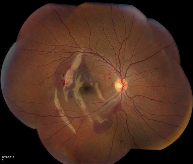
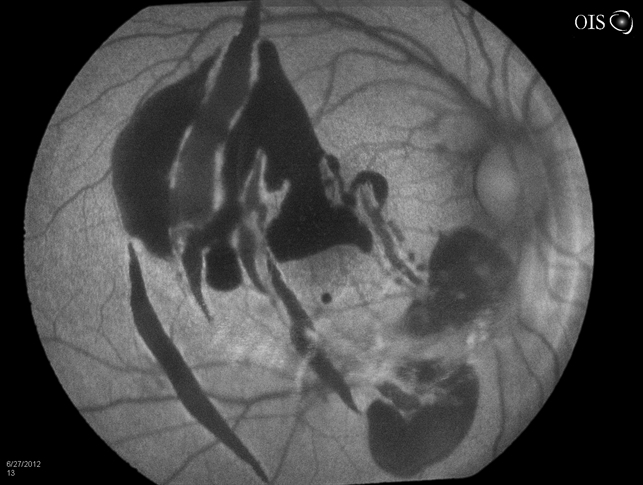
The findings were subretinal pigment epithelial hemorrhage and vertical choroidal rupture.
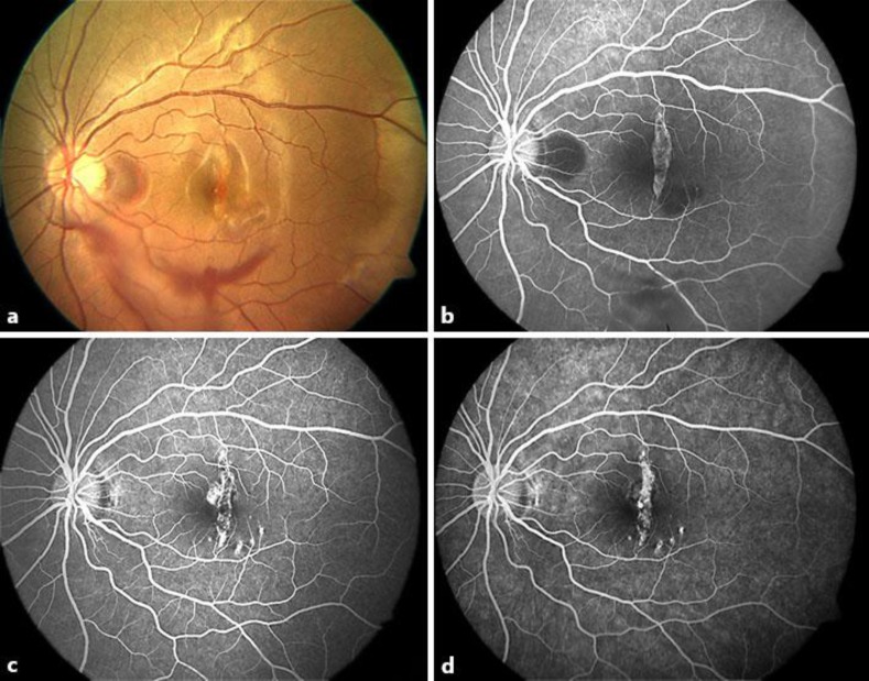
Three weeks after the trauma, the patient’s visual acuity was reduced to counting fingers at 30 cm.
On fundus examination and fluorescein angiography, subretinal pigment epithelial hemorrhage decreased, while the choroidal rupture expanded; on optical coherence tomography, outer retinal changes in the adjacent area were detected.
DISEASE
A choroidal rupture is a break in the choroid, Bruch membrane, and retinal pigment epithelium (RPE).
A choroidal rupture occurs as a result of the traumatic mechanical event directly at the site of contusion or more commonly away from the impact.
The force buckles the globe in at the area of impact and causes globe wall stress folding at a peripheral site causing the choroid, RPE, and Bruch membrane complex to stretch and break.
Fundus Explorer Pro
Photograph the retinal findings described here with the phone already in your pocket — 22 D optics and built-in illumination in one handheld unit.
From Choroida — the team behind this siteMANAGEMENT
Observation is recommended. An Amsler grid may be given to the patient and any changes in the grid should be reported to the patient’s eye care provider since a change may indicate the development of CNV.
However, in the presence of CNV, anti-vascular endothelial growth factor injections can be used.
There is no medical therapy available to cause the resolution of a choroidal rupture.


Document what you see
Two smartphone imaging tools built for everyday clinic use — one for the slit lamp, one for the fundus.
From Choroida — the team behind this siteComplications
Subretinal and subRPE hemorrhage can result at the time the choroidal rupture develops.
Choroidal neovascularization, usually type 2, can develop from the choroidal rupture where choroidal neovessels proliferate and grow in the subretinal space, leading to hemorrhage and fibrosis, and decreased vision if untreated.
Most CNV undergoes spontaneous resolution but many will need an anti-VEGF injection.
Test yourself
A few questions straight from this article.
-
A choroidal rupture is a break in which combination of structures?
A choroidal rupture is defined as a break in the choroid, Bruch membrane and the retinal pigment epithelium, which behave as one complex under stress. -
How does blunt trauma most commonly produce a choroidal rupture?
The force buckles the globe in at the area of impact and produces globe wall stress folding at a peripheral site, so ruptures more commonly arise away from the point of contact. -
What is the recommended management of a choroidal rupture with no neovascular complication?
Observation is recommended for an uncomplicated choroidal rupture, since no treatment shortens or reverses the break itself. -
Why is a patient with a choroidal rupture given an Amsler grid to use at home?
Any change the patient notices on the grid should be reported promptly, because it may indicate that choroidal neovascularization has developed. -
Which medical therapy causes a choroidal rupture itself to resolve?
The article is explicit that no medical therapy exists to cause resolution of a choroidal rupture; treatment is aimed only at its complications. -
Choroidal neovascularization arising from a choroidal rupture is usually of which type?
The neovascular complex is usually type 2: choroidal neovessels proliferate and grow within the subretinal space rather than remaining beneath the pigment epithelium. -
Which treatment is used when choroidal neovascularization complicates a choroidal rupture?
In the presence of choroidal neovascularization, anti-VEGF injections can be used; this is the one situation in which the article recommends active treatment. -
What is the usual natural course of choroidal neovascularization after a choroidal rupture?
Most of these neovascular membranes resolve spontaneously, although many patients still end up needing an anti-VEGF injection. -
Which complication can appear at the moment a choroidal rupture develops?
Subretinal and sub-retinal-pigment-epithelial haemorrhage may occur at the time the rupture forms, as in the reported case. -
In the reported case of choroidal rupture, how did the findings change during follow-up imaging?
On fundus examination and fluorescein angiography the sub-RPE haemorrhage decreased while the choroidal rupture itself expanded, and OCT showed outer retinal changes in the adjacent area.
