
Acute syphilitic posterior placoid chorioretinitis (ASPPC) is a specific fundus presentation of ocular syphilis, with a solitary or multifocal yellowish placoid lesion at the posterior pole that has a recognizable multimodal imaging signature.
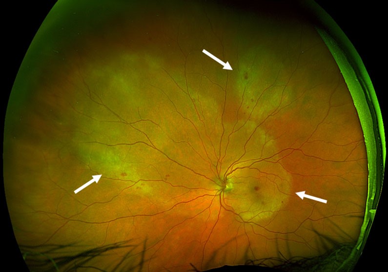
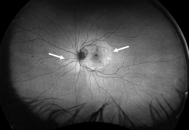
Acute syphilitic posterior placoid chorioretinitis is worth learning as its own pattern, because it is often mistaken for a primary inflammatory choroidopathy until syphilis serology comes back positive.
What Is Acute Syphilitic Posterior Placoid Chorioretinitis?
ASPPC is one of several posterior manifestations of ocular syphilis, alongside panuveitis, retinal vasculitis, and neuroretinitis (see ocular syphilis).
It typically occurs in the setting of secondary syphilis, and a substantial proportion of affected patients are HIV-positive; this does not change the ocular appearance, though it does affect systemic management and the urgency of neurosyphilis evaluation.
The lesion reflects inflammation at the level of the outer retina and RPE more than a primary choroidal process, which is why the imaging pattern differs from classic placoid choroiditis of other causes.
Clinical and Imaging Features
- A yellowish, placoid, usually round or oval lesion at the posterior pole, sometimes with a scalloped edge
- Mild to moderate vitritis in most eyes
- OCT shows disruption of the ellipsoid zone and outer retina overlying the lesion, with irregular thickening of the RPE-Bruch complex, sometimes described as a nodular pattern
- Fundus autofluorescence typically shows a granular hyperautofluorescent pattern within the lesion
- Fluorescein angiography shows early leopard-spot hypofluorescence with late staining, and indocyanine green angiography shows hypofluorescent spots
The combination of a placoid outer retinal lesion with vitritis in a sexually active adult should prompt syphilis testing even when other risk factors are not obvious.
Fundus Explorer Pro
Photograph the retinal findings described here with the phone already in your pocket — 22 D optics and built-in illumination in one handheld unit.
From Choroida — the team behind this siteDifferential Diagnosis
- Acute posterior multifocal placoid pigment epitheliopathy (APMPPE), which lacks vitritis and syphilis serology is negative (see APMPPE)
- Serpiginous choroidopathy, which is typically peripapillary and progressive, unlike the discrete placoid lesion of ASPPC (see serpiginous choroidopathy)
- Relentless placoid chorioretinitis, a rarer diffuse variant (see relentless placoid chorioretinitis)
- Tuberculous serpiginous-like choroiditis, more common in endemic regions
Serology settles the diagnosis in most cases, and treponemal tests remain positive for life, so a positive nontreponemal test with a compatible titer supports active or recent infection.
Workup
- Treponemal and nontreponemal serology (for example, FTA-ABS and RPR or VDRL)
- HIV testing, given the strong association
- Lumbar puncture to assess for neurosyphilis, since ocular syphilis is managed as a form of neurosyphilis regardless of cerebrospinal fluid findings
- Screening for other sexually transmitted infections as clinically indicated
Treatment
Ocular syphilis, including ASPPC, is treated with the neurosyphilis regimen: intravenous aqueous crystalline penicillin G for 10 to 14 days, or an alternative regimen for penicillin-allergic patients after desensitization when appropriate.
Vision often improves substantially once treatment starts, sometimes within days, and this rapid response is itself a useful diagnostic and prognostic sign.
Coordination with infectious disease specialists is standard, particularly for HIV-positive patients and for arranging follow-up serology to confirm treatment response.
Prognosis
Visual outcomes are generally favorable when penicillin therapy is started promptly, with many patients recovering good acuity even after significant initial vision loss.
Delayed diagnosis, often because the placoid appearance is mistaken for a primary inflammatory choroidopathy, is the main preventable cause of a worse outcome.


Document what you see
Two smartphone imaging tools built for everyday clinic use — one for the slit lamp, one for the fundus.
From Choroida — the team behind this siteReferences
- Eandi CM, Neri P, Adelman RA, Yannuzzi LA, Cunningham ET Jr; International Syphilis Study Group. Acute syphilitic posterior placoid chorioretinitis: report of a case series and comprehensive review of the literature. Retina. 2012;32:1915-1941.
- Pichi F, Ciardella AP, Cunningham ET Jr, et al. Spectral domain optical coherence tomography findings in patients with acute syphilitic posterior placoid chorioretinitis. Retina. 2014;34:373-384.
- Moradi A, Salek S, Daniel E, et al. Clinical features and incidence rates of ocular complications in patients with ocular syphilis. Am J Ophthalmol. 2015;159:334-343.
- Workowski KA, Bachmann LH, Chan PA, et al. Sexually transmitted infections treatment guidelines, 2021. MMWR Recomm Rep. 2021;70:1-187.
