
Periorbital ecchymosis — the common “black eye” — is bruising of the loose, vascular periorbital skin.
While the great majority of cases are exactly what they appear to be (a simple contusion from direct blunt trauma), the same finding is also one of the classic external signs of a basilar skull fracture, and telling these apart at the bedside is a genuinely important skill, because one requires nothing more than reassurance and cold compresses, and the other requires urgent neuroimaging.
The periorbital tissue bruises easily and dramatically because the skin there is thin and the subcutaneous space is loose, allowing blood to track and pool visibly even from a modest injury, which is part of why a black eye often looks more alarming than the underlying injury actually is.
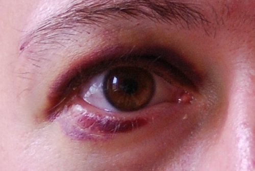
Simple Contusion
Direct blunt trauma to the periorbital region — a fall, a sports injury, an accidental elbow — causes local vessel rupture and bleeding into the subcutaneous tissue, which tracks under gravity and produces the characteristic discoloration that evolves through a predictable sequence of colors (red-purple, then blue-green, then yellow-brown) as the extravasated blood is broken down over one to two weeks.
Swelling typically peaks within the first 24 to 48 hours and then gradually resolves alongside the color changes, a predictable timeline that itself helps confirm a simple, uncomplicated contusion when the course matches expectations.
A simple contusion is usually isolated to the periocular region, develops promptly at the site of direct impact, and is not accompanied by other neurologic or ocular red flags.
Raccoon Eyes: Bilateral Periorbital Ecchymosis and Basilar Skull Fracture
Bilateral periorbital ecchymosis — “raccoon eyes” — occurring without a clear history of direct bilateral trauma is a recognized sign of anterior basilar skull fracture, where blood from the fracture site tracks forward along fascial planes into the periorbital tissue.
Unlike a simple contusion, this bruising often takes 1 to 3 days to develop after the initial head injury, because it depends on blood tracking a distance from the actual fracture site rather than appearing immediately at the point of impact.
This delayed onset is itself a useful clue: bruising that shows up a day or more after a head injury, especially when bilateral and not explained by direct trauma to both eyes, should raise concern for basilar skull fracture rather than being dismissed as a late-appearing simple bruise.
All-fit Slit-Lamp Adapter
Record and share exactly what you see at the slit lamp. One adapter fits any slit lamp or surgical microscope — and any smartphone.
From Choroida — the team behind this siteAssociated Findings Suggesting Basilar Skull Fracture
- Battle’s sign — bruising over the mastoid process behind the ear, from a middle cranial fossa fracture
- Cerebrospinal fluid rhinorrhea or otorrhea — clear fluid draining from the nose or ear, from a dural tear
- Hemotympanum — blood visible behind the tympanic membrane
- Cranial nerve deficits, particularly facial nerve palsy or hearing loss, from fracture lines crossing nerve canals
- Altered mental status or other signs of associated intracranial injury
None of these signs alone is required for the diagnosis; a basilar skull fracture can be present with bilateral periorbital ecchymosis as the only external clue, which is why any patient with a significant head injury and bilateral periorbital bruising warrants a careful search for the other findings above, performed deliberately rather than assumed absent simply because they are not immediately obvious.
Associated Ocular Injury
Because the same trauma that causes periorbital ecchymosis can also injure the globe or orbit, a periorbital bruise — regardless of whether it turns out to be simple or a marker of skull fracture — should prompt a full ocular exam: visual acuity, pupillary reaction, extraocular movements, and assessment for hyphema, globe rupture, or orbital fracture.
Reduced vision, a teardrop-shaped or irregular pupil, severe pain, or restricted eye movement are signs of a more serious associated ocular injury that changes the urgency of the workup considerably, and any of these findings should prompt urgent ophthalmologic evaluation rather than assuming the injury is limited to the periorbital soft tissue.
Evaluation
A history focused on mechanism, timing of bruise onset relative to any trauma, and associated symptoms (headache, vision change, drainage from nose or ear, hearing change) guides the decision to image, and this history is often more informative than the appearance of the bruise itself.
CT of the head and facial bones is the study of choice when basilar skull fracture, orbital fracture, or intracranial injury is a genuine concern, and should not be delayed in a patient with a significant mechanism of injury or any of the associated red flags above, even if the periorbital finding itself looks unremarkable.
Management
Simple periorbital contusion is managed conservatively: cold compresses in the first 24 to 48 hours to limit swelling, followed by warm compresses to encourage resorption, with resolution expected over one to two weeks without specific treatment.
Raccoon eyes from a confirmed basilar skull fracture are managed by treating the underlying fracture and any associated intracranial injury, typically with neurosurgical involvement, observation for cerebrospinal fluid leak, and prophylaxis against meningitis where indicated by the specific fracture pattern.
The ophthalmologist’s role in this scenario is often limited to confirming that the eye and orbit themselves have not sustained a separate, additional injury, while the broader head trauma is managed by the appropriate specialists.


Document what you see
Two smartphone imaging tools built for everyday clinic use — one for the slit lamp, one for the fundus.
From Choroida — the team behind this siteReferences
- Wall SP, Rice PL Jr. Periorbital ecchymosis and forensic considerations. Journal of Forensic Sciences.
- Bagheri N, Wajda B, eds. The Wills Eye Manual.
- Rhea JT, Rao PM. Helical CT and three-dimensional CT of facial and orbital injury. Radiologic Clinics of North America.
- American Academy of Ophthalmology. Basic and Clinical Science Course, Section 7: Orbit, Eyelids, and Lacrimal System.
Test yourself
A few questions straight from this article.
-
Why does periorbital tissue bruise so dramatically after a modest injury?
Thin periorbital skin over a loose subcutaneous space lets blood track and pool visibly, so a black eye often looks worse than the underlying injury really is. -
Through which colour sequence does a simple periorbital contusion evolve?
Extravasated blood is broken down over one to two weeks through this predictable colour sequence, and a course that matches it supports a simple contusion. -
How does blood reach the eyelids in raccoon eyes after a basilar skull fracture?
In anterior basilar skull fracture, blood from the fracture site tracks forward along fascial planes into periorbital tissue, producing bilateral bruising without direct injury there. -
How long after a head injury do raccoon eyes typically become apparent?
Because the blood must track a distance from the fracture, this bruising often takes 1 to 3 days to appear, and that delay is itself a diagnostic clue. -
Battle's sign refers to bruising in which location?
Battle's sign is mastoid bruising from a middle cranial fossa fracture, one of several external signs supporting a basilar skull fracture. -
A head-injured patient has bilateral periorbital bruising and no other external signs. What does this indicate?
A basilar skull fracture can present with bilateral periorbital ecchymosis as its only external clue, so the other associated signs must be searched for deliberately. -
Which finding after periorbital trauma signals a serious associated ocular injury?
Reduced vision, an irregular or teardrop pupil, severe pain or restricted movement point to globe or orbital injury and demand urgent ophthalmologic evaluation. -
Which imaging study is chosen when basilar or orbital fracture is a genuine concern?
CT of the head and facial bones is the study of choice and should not be delayed given a significant mechanism or red flags, even if the bruise itself looks unremarkable. -
How is a simple periorbital contusion managed?
Cold compresses in the first 24 to 48 hours limit swelling, then warm compresses encourage resorption, with resolution over one to two weeks and no specific treatment. -
How are raccoon eyes from a confirmed basilar skull fracture managed?
Treatment addresses the fracture and any intracranial injury, typically with neurosurgical involvement, observation for cerebrospinal fluid leak and meningitis prophylaxis where indicated.
