
CASE REPORT
A 32-year-old female patient presented to an ophthalmology clinic with complaints of difficulty in seeing in low light conditions, color vision problems, and decreased visual acuity.

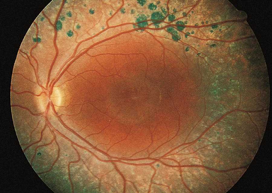
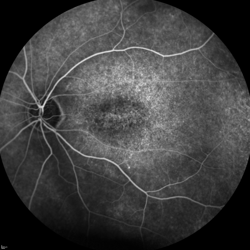
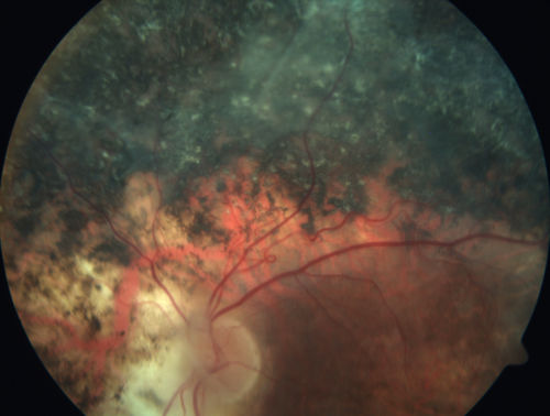
The patient’s medical and family history were unremarkable. Upon examination, her visual acuity was found to be 20/60 in both eyes. A fundus examination was performed, which revealed a pale optic disc, attenuated retinal vessels, and macular atrophy in both eyes.
An electroretinography (ERG) was performed, which showed an abnormal S-cone response in both eyes. Based on these findings, the patient was diagnosed with Enhanced S-Cone Syndrome (ESCS).
Enhanced S-Cone Syndrome DISEASE entity
Enhanced S Cone syndrome (ESCS) is a relatively newly described and very rare inherited progressive retinal degeneration that can cause severe vision loss. The characteristic electroretinogram (ERG) findings were first described in 1990 and are important in the diagnosis of this genetic disease.
Enhanced S Cone syndrome (ESCS) is a progressive autosomal recessive retinal degeneration caused by a mutation in nuclear receptor subfamily 2, group E, member 3 (NR2E3) gene, and rarely in the NRL gene.
The mutation of the NR2E3 receptor results in a gain of function in S cones. The S cone is one of three types of retinal cones and is the least common cone in the normal human retina. During embryology, the malfunctioning NR2E3 transcription factor is hypothesized to result in a differentiation error resulting in too many S cones being produced and no rods.
In rare cases, mutations in the NRL gene, which acts upstream of the NR2E3 transcription factor, can lead to ESCS.
Enhanced S-Cone Syndrome MANAGEMENT
Medical therapy
Macular schisis can be treated with oral or topical carbonic anhydrase inhibitors, although efficacy is variable.
Choroidal neovascularization can be treated with anti-vascular endothelial growth factor agents and has been used in children as young as 5 years.
Medical follow up
Children should be monitored for refractive error and macular status. OCT can be performed to monitor for schisis and choroidal neovascularization.
Would you have interest in taking retina images by smartphone?
Fundus photography is superior to fundus analysis as it enables intraocular pathologies to be photo-captured and encrypted information to be shared with colleagues and patients.
Recent technologies allow smartphone-based attachments and integrated lens adaptors to transform the smartphone into a portable fundus camera and Retinal imaging by smartphone.
RETINAL IMAGING BY YOUR SMARTPHONE
REFERENCES
- Jacobson SG, Marmor MF, Kemp CM, Knighton RW. SWS (blue) cone hypersensitivity in a newly identified retinal degeneration. Invest Ophthalmol Vis Sci. 1990;31(5):827-838.
- Haider NB, Mollema N, Gaule M, et al. Nr2e3-directed transcriptional regulation of genes involved in photoreceptor development and cell-type specific phototransduction. Exp Eye Res. 2009;89(3):365-372. doi:10.1016/j.exer.2009.04.006
- Littink KW, Stappers PTY, Riemslag FCC, et al. Autosomal Recessive NRL Mutations in Patients with Enhanced S-Cone Syndrome [published correction appears in Genes (Basel). 2018 Mar 07;9(3):]. Genes (Basel). 2018;9(2):68. Published 2018 Jan 30. doi:10.3390/genes9020068
- Tsang SH, Sharma T. Enhanced S-Cone Syndrome (Goldmann-Favre Syndrome). Adv Exp Med Biol. 2018;1085:153-156. doi: 10.1007/978-3-319-95046-4_28. PMID: 30578501.
- Khan, A. O., Aldahmesh, M., & Meyer, B. (2009). The enhanced S-cone syndrome in children. BMJ case reports, 2009, bcr10.2008.1163.
- de Carvalho ER, Robson AG, Arno G, Boon C, Webster AA, Michaelides M. Enhanced S-cone syndrome: spectrum of clinical, imaging, electrophysiological and genetic findings in a retrospective case series of 56 patients [published online ahead of print, 2020 Jul 14]. Ophthalmol Retina. 2020;S2468-6530(20)30286-4. doi:10.1016/j.oret.2020.07.008.
