
CASE REPORT
A 45-year-old male presented to an ophthalmology clinic with a chief complaint of visual disturbances and the presence of a central scotoma in his right eye.
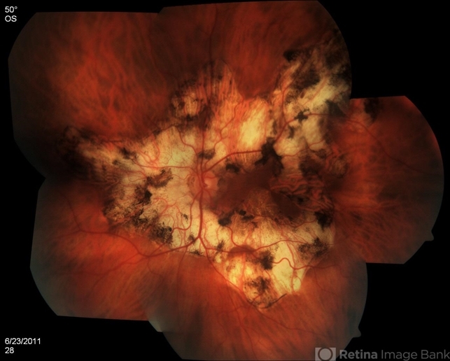
He reported a gradual decline in visual acuity over the past two months. Notably, there was no history of ocular trauma or systemic illnesses, including diabetes or hypertension.
On ocular examination, the visual acuity in the right eye (OD) was measured at 20/200, while the left eye (OS) exhibited 20/20 vision.
Intraocular pressure measurements were within the normal range, with OD at 15 mmHg and OS at 16 mmHg.
In the posterior segment examination of the right eye (OD), a central scotoma was observed during Amsler grid testing.
Additionally, mild vitritis was noted along with the presence of multiple yellow-white, deep choroidal lesions at the posterior pole, extending into the macular region.
This constellation of findings was consistent with active choroiditis and areas of serpiginous geographic atrophy. In contrast, the left eye (OS) demonstrated a normal retina and choroid.
Diagnostic tests, including fluorescein angiography, indicated hyperfluorescence of the choroidal lesions in the late phase, with no evidence of active neovascularization. Optical coherence tomography further revealed subretinal fluid accumulation and outer retinal disruption in the macular area.
Based on the clinical presentation, ophthalmic examination, and diagnostic test results, the patient was diagnosed with Serpiginous Choroidopathy (SC).
Serpiginous Choroidopathy DISEASE entity
Serpiginous choroidopathy (SC) is a rare, bilateral, chronic, progressive, recurrent inflammatory disease of the retinal pigment epithelium (RPE), choriocapillaris, and choroid of unknown etiology.
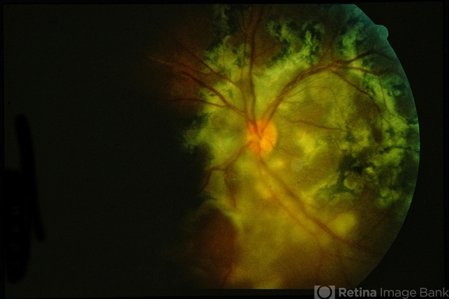
SC is a rare clinical entity causing less than 5% of posterior uveitis cases. It has a higher prevalence in men and affects young to middle-aged adults. No systemic disease associations have been identified.
Patients present with painless unilateral vision loss, metamorphosis, or central scotoma.
Although of unknown etiology, its origin is probably immunogenic since it seems to respond to treatment with corticosteroids and other immunosuppressants. Moreover, affected patients also show an increased frequency of HLA-B7 and retinal S-antigen associations.
Other pathogenic mechanisms have been proposed and there may be an association with SC with infectious etiologies such as Mycobacterium tuberculosis and herpes viruses but this remains to be proven. Alternatively, SC and tuberculosis-related SC may be different disease entities with overlapping features.
Fundus Explorer Pro
Photograph the retinal findings described here with the phone already in your pocket — 22 D optics and built-in illumination in one handheld unit.
From Choroida — the team behind this siteSerpiginous Choroidopathy Diagnosis
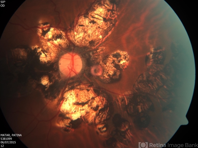
Serpiginous choroidopathy (SC) presents with gray-yellowish subretinal infiltrates that usually spread centrifugally from the peripapillary region in a serpiginous (snake-like) manner. Active lesions show a leading edge and resolve with subsequent RPE and choriocapillary atrophy.
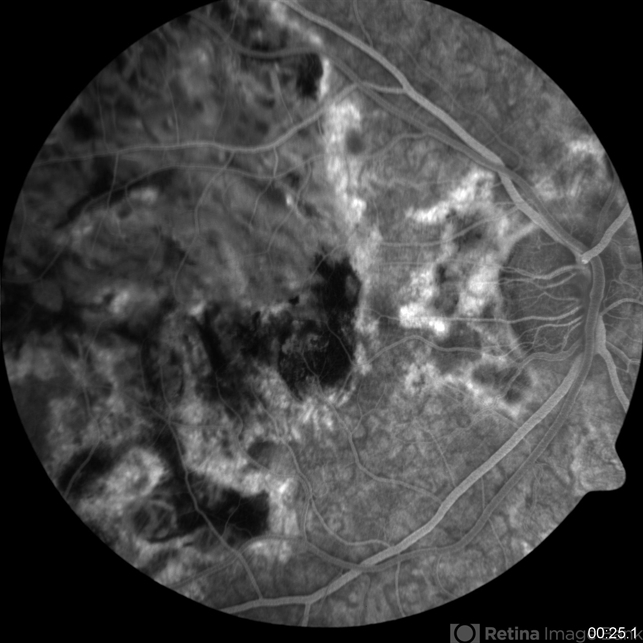
Consecutive recurrences cause further atrophy leaving hypo- and hyper-pigmented lesions that spread irregularly over the posterior fundus. Although bilateral, the disease is often asymmetric with multiple lesions in different stages of resolution in both eyes.
Recurrences have variable intervals that range from months to years. Anterior chamber and vitritis are minimal.
The former begins as a macular lesion that spares the peripapillary region with a higher risk of choroidal neovascularization (CNV) and poor visual outcome.
Ampiginous chorioretinopathy is characterized by multifocal plaque-like lesions scattered over the posterior pole that have overlapping features of both acute posterior multifocal placoid pigment epitheliopathy (APMPPE) and SC.
Ampiginous chorioretinopathy is characterized by multiple recurrences and progressive enlargement of serpiginous lesions over time without the spontaneous resolution typical of APMPPE.
Serpiginous Choroidopathy MANAGEMENT
Treatment of Serpiginous choroidopathy (SC) aims to stop chorioretinal inflammation especially when the advancing lesions threaten the fovea.
Systemic or periocular corticosteroids are frequently used but recurrence prevention usually requires long-term anti-inflammatory treatment with a steroid-sparing agent, such as antimetabolites (methotrexate, mycophenolate mofetil, azathioprine), cyclosporine A, or anti-TNF agents, such as adalimumab or infliximab, once tuberculosis has been ruled out.
Long-term management can be challenging and up to 25% of the eyes have a final visual acuity of less than 20/200.
Complications
The most common complication of SC is choroidal neovascularization affecting up to 35% of patients. Other reported complications are subretinal fibrosis, cystoid macular edema, branch vein occlusion, serous retinal detachment, optic disc neovascularization, and anterior uveitis.


Document what you see
Two smartphone imaging tools built for everyday clinic use — one for the slit lamp, one for the fundus.
From Choroida — the team behind this siteREFERENCES
- Bacin F, Larmande J, Boulmier A, Juliiar G. Serpiginous choroiditis and placoid epitheliopathy. Bull Soc Ophthalmol Fr. 1983; 83:1153-62.
- Abu el-Asrar AM. Serpiginous (geographical) choroiditis. Int Ophthalmol Clin. 1995; 35:87-91
- Ciulla TA, Gragoudas ES. Serpiginous choroiditis. Int Ophthalmol Clin. 1996; 36:135-43.
- Nazari Khanamiri H, Rao NA. Serpiginous choroiditis and infectious multifocal serpiginoid choroiditis. Surv Ophthalmol. 2013 May-Jun;58(3):203-32.
- Gupta V, Gupta A, Arora S, Bambery P, Dogra MR, Agarwal A. Presumed tubercular serpiginous like choroiditis: clinical presentations and management. Ophthalmology. 2003 Sep;110(9):1744-9.
Test yourself
A few questions straight from this article.
-
Which tissues are primarily involved in serpiginous choroidopathy?
Serpiginous choroidopathy is a chronic, progressive, recurrent inflammatory disease of the retinal pigment epithelium, choriocapillaris and choroid, of unknown cause. -
What share of posterior uveitis cases is attributable to serpiginous choroidopathy?
Serpiginous choroidopathy is a rare entity accounting for under 5% of posterior uveitis, with a higher prevalence in men and no systemic disease association. -
Which immunological associations are increased in patients with serpiginous choroidopathy?
An immunogenic origin is suspected because affected patients show increased frequency of HLA-B7 and retinal S-antigen associations, and the disease responds to immunosuppression. -
How do the subretinal infiltrates of serpiginous choroidopathy characteristically spread?
Grey-yellowish subretinal infiltrates usually spread outward from the peripapillary region in a snake-like, serpiginous pattern. -
What happens to active serpiginous choroidopathy lesions as they resolve?
Active lesions display a leading edge and heal with retinal pigment epithelial and choriocapillary atrophy; repeated recurrences leave hypo- and hyperpigmented scarring. -
Which feature separates ampiginous chorioretinopathy from acute posterior multifocal placoid pigment epitheliopathy?
Ampiginous chorioretinopathy shares features with both conditions but shows multiple recurrences and progressive enlargement rather than the spontaneous resolution seen in placoid epitheliopathy. -
Over what interval do recurrences of serpiginous choroidopathy typically occur?
Recurrences occur at variable intervals ranging from months to years, leaving lesions at different stages of resolution in the two eyes. -
Before starting anti-TNF therapy such as adalimumab for serpiginous choroidopathy, what must be excluded?
Long-term steroid-sparing therapy may include anti-TNF agents such as adalimumab or infliximab, but only once tuberculosis has been ruled out. -
What is the most common complication of serpiginous choroidopathy?
Choroidal neovascularisation is the commonest complication, affecting up to 35% of patients, ahead of subretinal fibrosis and cystoid macular oedema. -
What proportion of eyes with serpiginous choroidopathy end with visual acuity worse than 20/200?
Long-term management is difficult, and up to a quarter of affected eyes finish with a visual acuity of less than 20/200.
