
Case Study
A 32-year-old female presented to an ophthalmology clinic with complaints of progressive blurred vision and photophobia in both eyes over the past month.

She had no significant medical history and denied any recent infections or trauma. The patient reported that her symptoms started with mild visual disturbances, which rapidly worsened.
Examination Results:
- Visual Acuity: 20/40 in both eyes.
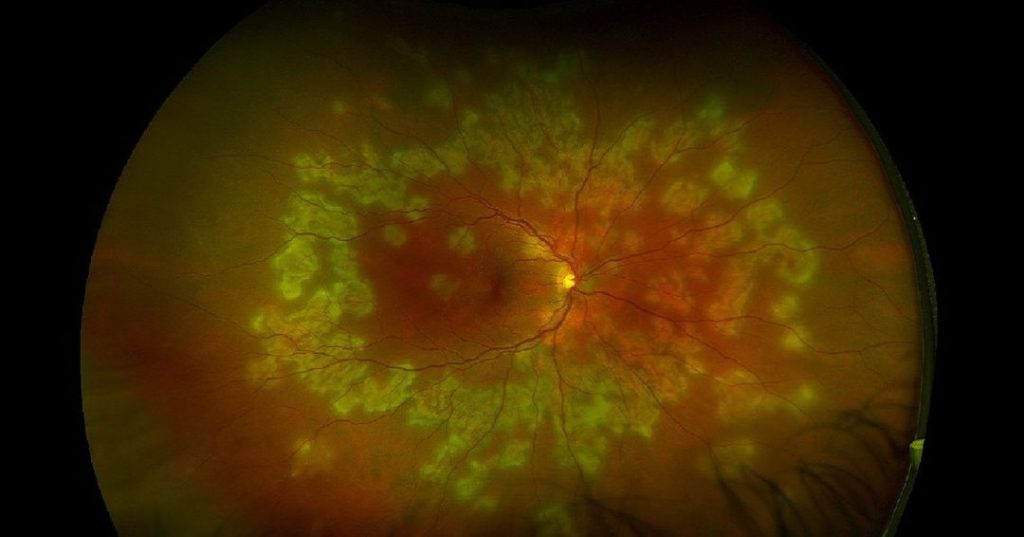
- Fundus Examination: Multiple large, creamy yellow placoid lesions in the posterior pole extending to the mid-periphery in both eyes.
- Fluorescein Angiography: Early hypofluorescence with late hyperfluorescence of the placoid lesions.
- Optical Coherence Tomography (OCT): Hyperreflective lesions at the level of the retinal pigment epithelium (RPE) with overlying retinal edema.
- Blood Work: Normal complete blood count (CBC) and erythrocyte sedimentation rate (ESR).
A diagnosis of Relentless Placoid Chorioretinitis (RPC) was confirmed.
Relentless Placoid Chorioretinitis (RPC) Disease entity
Relentless Placoid Chorioretinitis (RPC) is a rare, bilateral disease of the retinal pigment epithelium (RPE) and choroid, first described by Jones et al.
Due to the similarities of acute lesions with serpiginous choroiditis (SC) and acute posterior multifocal placoid pigment epitheliopathy (APMPPE), the diagnosis of Relentless Placoid Chorioretinitis (RPC) is often delayed due to disease overlap.
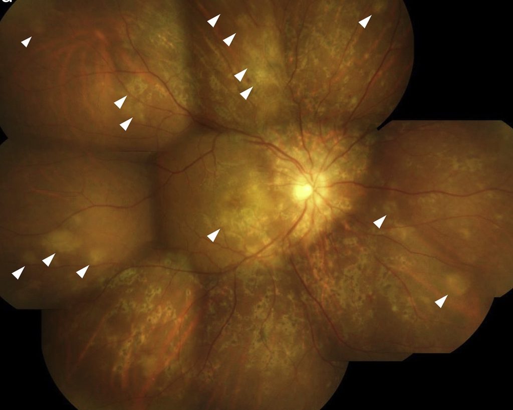
The distinction of RPC consists of its atypical time course and retinal distribution. RPC is characterized by >50 bilateral chorioretinal lesions with involvement anterior and posterior to the equator.
This is in contrast to SC, where, usually, only one eye contains active lesions at a time, consisting of subretinal infiltrates that spread centrifugally from the peripapillary region and result in a typical geographic scar.
Similar to SC, however, the clinical course of RPC is prolonged and relapsing, with new lesions continuing to appear for 5–24 months without therapy. Patients may show recurrences for months to years after onset.
Fundus Explorer Pro
Photograph the retinal findings described here with the phone already in your pocket — 22 D optics and built-in illumination in one handheld unit.
From Choroida — the team behind this sitePathophysiology
The exact pathophysiology of Relentless Placoid Chorioretinitis (RPC) remains unclear, but it is believed to be an immune-mediated inflammatory response targeting the retina and choroid.
The inflammatory process involves the deposition of immune complexes and subsequent damage to the retinal pigment epithelium (RPE) and choroidal vasculature.
This leads to the formation of the characteristic placoid lesions and retinal edema. The relentless nature of the condition suggests a continuous or recurrent inflammatory stimulus.
Epidemiology
Relentless Placoid Chorioretinitis (RPC) is a rare condition with limited epidemiological data available.
It typically affects young to middle-aged adults and has no clear gender or racial predilection.
The rarity of the condition and its variable presentation make it challenging to diagnose and study, contributing to the limited understanding of its epidemiology.
Relentless Placoid Chorioretinitis (RPC) Clinical Features
Patients with Relentless Placoid Chorioretinitis (RPC) often present with a range of symptoms that reflect the extent and progression of the inflammatory process:
- Blurred Vision: A common initial symptom due to retinal and choroidal involvement.
- Photophobia: Sensitivity to light caused by inflammation and retinal irritation.
- Visual Field Defects: Peripheral vision loss or scotomas corresponding to the location of the placoid lesions.
- Floaters: Resulting from vitreous involvement in some cases.
Examination Findings
- Fundus Examination: Reveals multiple large, creamy yellow placoid lesions in the posterior pole and mid-periphery of both eyes.
- Fluorescein Angiography: Shows early hypofluorescence with late hyperfluorescence of the placoid lesions, indicating active inflammation and leakage.
- Optical Coherence Tomography (OCT): Displays hyperreflective lesions at the RPE level with overlying retinal edema.
- Systemic Evaluation: Typically unremarkable, although comprehensive systemic workup is recommended to rule out infectious or systemic inflammatory conditions.

Relentless Placoid Chorioretinitis (RPC) Differential Diagnosis
The differential diagnosis for Relentless Placoid Chorioretinitis (RPC) includes several other inflammatory and infectious conditions with similar clinical features:
- Acute Posterior Multifocal Placoid Pigment Epitheliopathy (APMPPE): Characterized by multiple placoid lesions but typically self-limited and less aggressive.
- Serpiginous Choroiditis: Presents with serpentine lesions and chronic, recurrent inflammation.
- Syphilitic Chorioretinitis: Can present with placoid lesions but usually associated with systemic signs of syphilis.
- Viral Retinitis: Such as cytomegalovirus (CMV) retinitis, which presents with retinal necrosis and hemorrhages.
Relentless Placoid Chorioretinitis (RPC) Diagnosis
Diagnosing Relentless Placoid Chorioretinitis (RPC) involves a combination of clinical evaluation and imaging studies:
- Clinical Examination: Detailed fundus examination to identify characteristic placoid lesions.
- Fluorescein Angiography: Essential for visualizing the pattern of early hypofluorescence and late hyperfluorescence.
- Optical Coherence Tomography (OCT): Provides detailed imaging of retinal structures, highlighting lesions at the RPE level and associated edema.
- Systemic Workup: Includes tests for infectious and systemic inflammatory conditions to rule out other causes.
Relentless Placoid Chorioretinitis (RPC) Management
Management of Relentless Placoid Chorioretinitis (RPC) focuses on controlling the inflammatory process and preventing further retinal damage:
- Corticosteroids: High-dose oral or intravenous corticosteroids are the mainstay of treatment to reduce inflammation.
- Immunosuppressive Agents: Such as azathioprine or mycophenolate mofetil may be used in refractory cases or to reduce steroid dependence.
- Intravitreal Injections: Anti-VEGF agents or corticosteroids may be considered for persistent macular edema.
- Regular Monitoring: Frequent follow-up to monitor disease progression and treatment response.
Prognosis
The prognosis for patients with Relentless Placoid Chorioretinitis (RPC) varies depending on the severity of inflammation and the effectiveness of treatment.
Early diagnosis and aggressive management can significantly improve outcomes. However, chronic or recurrent inflammation can lead to permanent retinal damage and visual impairment in some cases.
Prevention
There are no specific preventive measures for Relentless Placoid Chorioretinitis (RPC) due to its unknown etiology.
Public health efforts should focus on raising awareness among healthcare providers to ensure timely diagnosis and management.
Early intervention can help prevent significant visual impairment and improve outcomes for patients with Relentless Placoid Chorioretinitis (RPC).


Document what you see
Two smartphone imaging tools built for everyday clinic use — one for the slit lamp, one for the fundus.
From Choroida — the team behind this siteReferences
- Thorne, J. E., Wittenberg, S., Jabs, D. A., Peters, G. B., & Dunn, J. P. (2006). Multifocal choroiditis with panuveitis: incidence of ocular complications and of loss of visual acuity. Ophthalmology, 113(12), 2310-2316.
- Yannuzzi, L. A., Wong, D. W., Sforzolini, B. S., Goldbaum, M., & Smith, R. T. (2000). Relentless placoid chorioretinitis: a new entity or an unusual variant of serpiginous choroiditis?. Archives of Ophthalmology, 118(6), 931-938.
- Abu El-Asrar, A. M., Herbort, C. P., & Tabbara, K. F. (2010). Serpiginous choroiditis. Ocular Immunology and Inflammation, 18(5), 395-403.
- Gaudio, P. A. (2004). Inflammatory diseases of the retina and vitreous. Retina, 3, 1631-1643.
- Foster, C. S., & Vitale, A. T. (2002). Diagnosis and Treatment of Uveitis. W.B. Saunders Company.
Test yourself
A few questions straight from this article.
-
Relentless placoid chorioretinitis is a rare disease of which structures?
Relentless placoid chorioretinitis is a rare bilateral disease of the retinal pigment epithelium and the choroid, and its acute lesions resemble those of serpiginous choroiditis and APMPPE. -
Which lesion burden characterises relentless placoid chorioretinitis?
What distinguishes relentless placoid chorioretinitis is its retinal distribution: more than 50 bilateral chorioretinal lesions, with involvement both anterior and posterior to the equator. -
For how long do new lesions keep appearing in untreated relentless placoid chorioretinitis?
The course is prolonged and relapsing: without therapy new lesions continue to appear for 5 to 24 months, and recurrences may occur for months to years after onset. -
How does serpiginous choroiditis differ from relentless placoid chorioretinitis in its pattern of activity?
In serpiginous choroiditis usually only one eye has active lesions at a time: subretinal infiltrates spread centrifugally from the peripapillary region and leave a typical geographic scar. -
How does acute posterior multifocal placoid pigment epitheliopathy differ from relentless placoid chorioretinitis?
APMPPE also shows multiple placoid lesions, but it is typically self-limited and less aggressive than relentless placoid chorioretinitis, whose course is prolonged and relapsing. -
What mechanism is believed to underlie relentless placoid chorioretinitis?
The disease is thought to be an immune-mediated inflammatory response in which immune complex deposition damages the retinal pigment epithelium and the choroidal vasculature, producing the placoid lesions and retinal oedema. -
What do the fundus lesions of relentless placoid chorioretinitis look like?
Fundus examination shows multiple large, creamy yellow placoid lesions in the posterior pole and mid-periphery of both eyes. -
What fluorescein angiographic pattern do the placoid lesions of relentless placoid chorioretinitis show?
The lesions show early hypofluorescence followed by late hyperfluorescence, a pattern that indicates active inflammation and leakage. -
Why is a comprehensive systemic workup recommended when relentless placoid chorioretinitis is suspected?
Systemic evaluation is typically unremarkable in this disease, but a comprehensive workup is recommended to exclude infectious and systemic inflammatory conditions such as syphilitic chorioretinitis, which can also produce placoid lesions. -
What is the mainstay of treatment for relentless placoid chorioretinitis?
High-dose oral or intravenous corticosteroids are the mainstay of treatment; immunosuppressive agents such as azathioprine or mycophenolate mofetil are added in refractory cases or to reduce steroid dependence.
