
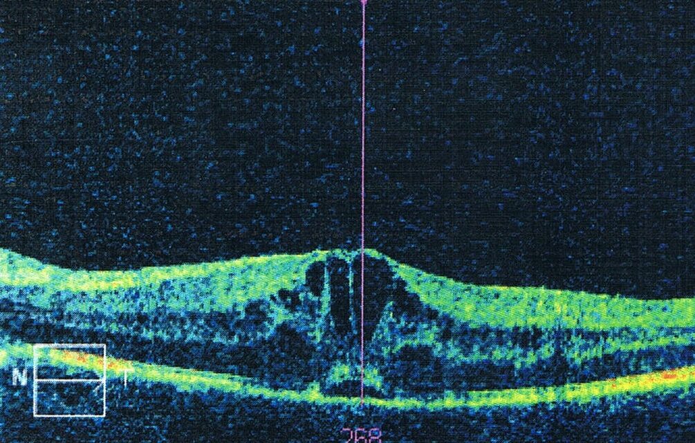
Irvine-Gass syndrome is cystoid macular edema that develops in the weeks after cataract surgery, and it is the most common cause of unexpected poor vision after an otherwise uneventful operation.
It was described by Irvine in 1953 and characterized angiographically by Gass and Norton in 1966.
Most cases resolve with treatment or on their own, though a minority become chronic and leave lasting visual loss.
Recognizing the condition early, and knowing who is at higher risk, allows targeted prophylaxis and prompt treatment.
What Is Irvine-Gass Syndrome?
The term describes pseudophakic cystoid macular edema (PCME), meaning macular edema after cataract surgery without another explanation such as diabetes or vein occlusion.
Onset is typically four to twelve weeks after surgery, with a peak around six weeks.
Angiographic leakage is common, but clinically significant edema with reduced vision occurs in a small percentage of eyes after uncomplicated phacoemulsification.
OCT-detected subclinical thickening is more frequent, and many of these eyes never develop symptoms.
Pathogenesis
Surgical trauma and inflammation release prostaglandins and other inflammatory mediators into the anterior and posterior segment.
These mediators increase permeability of the perifoveal capillaries and cause fluid to accumulate in the outer plexiform and inner nuclear layers.
Vitreous traction on the macula, or vitreous incarcerated in the wound, may contribute in some eyes.
Topical prostaglandin analogues may increase the risk in susceptible eyes.
Light toxicity from the operating microscope has also been proposed as a minor contributor.
Fundus Explorer Pro
Photograph the retinal findings described here with the phone already in your pocket — 22 D optics and built-in illumination in one handheld unit.
From Choroida — the team behind this siteRisk Factors
- Diabetes mellitus, with or without retinopathy
- Uveitis and chronic inflammation
- Epiretinal membrane
- Prior retinal vein occlusion
- History of CME after surgery in the fellow eye
- Posterior capsule rupture and vitreous loss
- Retained lens fragments and iris trauma
- Complicated surgery with prolonged operative time
- Topical prostaglandin analogue use
- Age-related macular degeneration and hypertension in some studies
A large database study found that diabetes and uveitis, as well as prior CME in the other eye and vitreous loss, were among the strongest predictors of edema.
Clinical Presentation
Patients report blurred or distorted central vision that follows an initial period of good recovery.
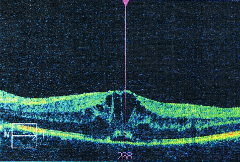
Examination shows a pseudophakic eye with a normal or mildly inflamed anterior segment, and the macula appears thickened with a loss of the foveal reflex and cystic changes.
Vitreous cells may be present.
Other causes of poor vision after surgery should be considered, including posterior capsule opacification, IOL malposition, retinal detachment, and endophthalmitis.
Diagnosis and Imaging
OCT
OCT is the standard test.
- Cystoid spaces in the outer plexiform and inner nuclear layers, with central thickening
- Subretinal fluid in some eyes
- Vitreomacular traction or epiretinal membrane when present (see vitreomacular traction)
Fluorescein Angiography
Angiography shows the petaloid leakage pattern typical of cystoid edema and often disc hyperfluorescence, which is a characteristic feature of Irvine-Gass syndrome.
It is not always needed, but it helps in unclear cases.
Assessment of the Anterior Segment
Slit-lamp examination checks for iris prolapse, vitreous strands in the wound, or malpositioned IOL haptics, which may cause chronic irritation (see UGH syndrome).
Differential Diagnosis
- Diabetic macular edema, which may coexist or be unmasked by surgery
- Retinal vein occlusion
- Uveitic macular edema
- Epiretinal membrane
- Neovascular AMD
- Drug-induced macular edema
Pre-existing macular disease should be excluded by preoperative OCT in patients at risk.
Prevention
Topical NSAIDs are widely used around cataract surgery to reduce prostaglandin-mediated inflammation.
The ESCRS PREMED study evaluated topical treatments in both diabetic and nondiabetic patients, and the combination of a topical NSAID with a steroid gave better protection against macular thickening than a steroid alone, particularly in diabetic patients.
A practical approach includes:
- Preoperative NSAID in high-risk patients, continuing for several weeks after surgery
- Careful surgical technique to minimize trauma and vitreous loss
- Preoperative OCT in patients with diabetes, uveitis, or macular disease
- Control of inflammation before surgery in uveitic eyes, ideally for at least three months
- Monitoring for high-risk patients at four to six weeks after surgery
Treatment
Topical Therapy
The first-line treatment combines a topical NSAID, such as ketorolac, nepafenac, or bromfenac, with a topical corticosteroid, such as prednisolone acetate.
Treatment continues for several weeks, with review by OCT.
Regional and Intravitreal Corticosteroids
Periocular triamcinolone, intravitreal triamcinolone, or a dexamethasone implant is used when topical therapy fails (see role of steroids in resistant edema).
Steroids raise IOP and are used with monitoring.
Other Options
- Oral or topical carbonic anhydrase inhibitors
- Intravitreal anti-VEGF injection, with variable results
- YAG vitreolysis to cut vitreous strands in the wound
- Vitrectomy for chronic edema with vitreous traction or incarceration
Natural History
Most cases resolve within three to twelve months, whether or not treated, and a minority persist as chronic edema with photoreceptor damage.
Early treatment shortens the course and prevents chronic changes.
Prognosis
The visual prognosis is good in most patients.
Chronic edema over many months reduces final acuity, and eyes with retinal or vascular comorbidity do worse.
Patients should be informed that recovery may take months, and that vision may improve slowly after the fluid resolves.
See also the general review of cystoid macular edema (see cystoid macular edema).


Document what you see
Two smartphone imaging tools built for everyday clinic use — one for the slit lamp, one for the fundus.
From Choroida — the team behind this siteReferences
- Irvine SR. A newly defined vitreous syndrome following cataract surgery, interpreted according to recent concepts of the structure of the vitreous. Am J Ophthalmol. 1953;36:599-619.
- Gass JDM, Norton EW. Cystoid macular edema and papilledema following cataract extraction: a fluorescein fundoscopic and angiographic study. Arch Ophthalmol. 1966;76:646-661.
- Chu CJ, Johnston RL, Buscombe C, Sallam AB, Mohamed Q, Yang YC; UK Pseudophakic Macular Edema Study Group. Risk factors and incidence of macular edema after cataract surgery: a database study of 81984 eyes. Ophthalmology. 2016;123:316-323.
- Henderson BA, Kim JY, Ament CS, Ferrufino-Ponce ZK, Grabowska A, Cremers SL. Clinical pseudophakic cystoid macular edema: risk factors for development and duration after treatment. J Cataract Refract Surg. 2007;33:1550-1558.
- Wielders LHP, Schouten JSAG, Winkens B, et al. European multicenter trial of the prevention of cystoid macular edema after cataract surgery in nondiabetics: ESCRS PREMED study report 1. J Cataract Refract Surg. 2018;44:429-439.
- Wielders LHP, Schouten JSAG, Winkens B, et al. Randomized controlled European multicenter trial on the prevention of cystoid macular edema after cataract surgery in diabetics: ESCRS PREMED study report 2. J Cataract Refract Surg. 2018;44:836-847.
