
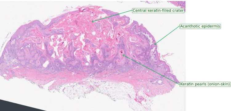
Keratoacanthoma is a rapidly growing, dome-shaped epidermal tumor with a characteristic central keratin-filled crater that, on the eyelid as elsewhere on sun-exposed skin, presents a genuine diagnostic puzzle: it behaves like a benign, self-limited lesion usually, yet it is histologically difficult to distinguish from well-differentiated squamous cell carcinoma.
There remains debate about whether it should be considered a distinct entity or a variant of SCC, and this unresolved classification question has direct, practical consequences for how confidently a clinician can rely on the “wait and watch” approach sometimes used elsewhere on the body.
On the eyelid specifically, the stakes of getting this distinction wrong are higher than on typical trunk or limb skin, given the risk to both vision and the globe from local tissue destruction, and because the confined, functionally critical anatomy of the lid leaves far less margin for a delayed or incorrect diagnosis than more forgiving skin elsewhere on the body.
Clinical Presentation
The hallmark of keratoacanthoma is its growth pattern: a firm, dome-shaped, flesh-colored to erythematous nodule with a central keratin plug appears and grows rapidly — often reaching significant size within just four to eight weeks — a tempo that is distinctly faster than the slow, indolent growth typical of basal cell or squamous cell carcinoma.
This rapid growth phase is followed, in the classic natural history, by a period of stability and then, if left untreated, spontaneous involution over several months, leaving a depressed scar.
On the eyelid, this rapid growth carries specific risks beyond the cosmetic scarring seen elsewhere: significant lid margin distortion, lash loss, mechanical ptosis from lesion bulk, or exposure keratopathy if the growing lesion interferes with normal eyelid closure.
Because these functional consequences can develop within the same few weeks the lesion itself is rapidly enlarging, watching and waiting to confirm the diagnosis clinically carries a real, tangible cost on the eyelid that it would not carry at a less functionally critical site.
Risk Factors
- Chronic sun exposure — the dominant risk factor, consistent with its typical location on sun-exposed skin including the eyelids
- Fair skin and older age
- Immunosuppression, including solid organ transplant recipients, in whom keratoacanthomas can be more numerous and more likely to behave aggressively
- Prior trauma or surgical scar at the site, in some reported cases
- Certain genetic syndromes (Muir-Torre syndrome, Ferguson-Smith syndrome) predisposing to multiple keratoacanthomas
All-fit Slit-Lamp Adapter
Record and share exactly what you see at the slit lamp. One adapter fits any slit lamp or surgical microscope — and any smartphone.
From Choroida — the team behind this siteDifferential Diagnosis
- Squamous cell carcinoma — the most important alternative diagnosis, given the histologic overlap and the fact that some lesions initially called keratoacanthoma are later reclassified
- Basal cell carcinoma — typically slower growing, pearly rolled border, less likely to have the central keratin plug
- Molluscum contagiosum — smaller, umbilicated, often multiple, more common in children or immunosuppressed adults
- Cutaneous horn — a descriptive term for a keratin projection that can overlie several different underlying lesions, including keratoacanthoma
- Chalazion or hordeolum — inflammatory, softer, without the characteristic central crater
Because the rapid-growth-then-involution pattern is only reliably recognized in retrospect, and because the histologic distinction from SCC is difficult even for experienced pathologists, most clinicians treat a suspected eyelid keratoacanthoma with the same seriousness and the same excisional approach used for a confirmed malignancy, rather than relying on the possibility of spontaneous resolution.
Diagnostic Evaluation
Excisional biopsy, rather than a small incisional or shave biopsy, is generally preferred when feasible, because it provides the pathologist with the full architecture of the lesion (which is important for distinguishing keratoacanthoma from SCC) and is simultaneously therapeutic.
When the clinical appearance and growth pattern are classic and biopsy is being deferred, close short-interval follow-up is essential to confirm the lesion is following the expected benign course rather than continuing to enlarge.
Management
Complete surgical excision with clear margins is the standard treatment for eyelid keratoacanthoma, given both the diagnostic uncertainty relative to squamous cell carcinoma and the anatomic risks of allowing a rapidly growing lesion to progress unchecked in this location.
Eyelid reconstruction techniques are often needed to restore both function and cosmesis after excision, for larger lesions involving a significant portion of the lid margin, following the same general reconstructive principles used for other eyelid tumors requiring wide local excision.
Observation for spontaneous involution — sometimes considered for classic, small, non-eyelid keratoacanthomas — is generally not favored on the eyelid, given the risk of significant local tissue destruction, scarring, and lid margin disruption during the growth phase, even if the lesion would eventually resolve on its own.
Prognosis
With complete excision, prognosis is excellent and recurrence is uncommon, and most patients experience no lasting functional consequence once the eyelid has been appropriately reconstructed.
Any lesion with atypical features, incomplete excision, or histology that raises genuine concern for squamous cell carcinoma should be managed with the same follow-up and, where appropriate, referral pathways used for confirmed eyelid SCC, given the overlapping biology between the two.
Given this diagnostic overlap, communication between the surgeon and pathologist matters more than usual for this particular lesion, since the final diagnosis and the resulting management plan often depend on a genuinely collaborative interpretation of both the clinical growth pattern and the histologic findings together, rather than either being fully decisive on its own.


Document what you see
Two smartphone imaging tools built for everyday clinic use — one for the slit lamp, one for the fundus.
From Choroida — the team behind this siteReferences
- Kwiek B, Schwartz RA. Keratoacanthoma (KA): an update and review. Journal of the American Academy of Dermatology.
- Savar A, Oellers P, Myers J, et al. Positive predictive value of lymphoscintigraphy and sentinel lymph node biopsy in eyelid and conjunctival malignancy. Ophthalmic Plastic and Reconstructive Surgery.
- American Academy of Ophthalmology. Basic and Clinical Science Course, Section 7: Orbit, Eyelids, and Lacrimal System.
- Cook BE Jr, Bartley GB. Treatment options and future prospects for the management of eyelid malignancies. Ophthalmology.
Test yourself
A few questions straight from this article.
-
Which morphology is the hallmark of an eyelid keratoacanthoma?
Keratoacanthoma is a firm, dome-shaped, flesh-coloured to erythematous nodule with a characteristic central keratin-filled crater. -
Over what period does an eyelid keratoacanthoma typically reach significant size?
Keratoacanthoma often reaches significant size within four to eight weeks, a tempo distinctly faster than basal or squamous cell carcinoma. -
In its classic natural history, what happens to an untreated keratoacanthoma after its growth phase?
Rapid growth is followed by a period of stability, then spontaneous involution over several months, leaving a depressed scar. -
What is the dominant risk factor for eyelid keratoacanthoma?
Chronic sun exposure is the dominant risk factor, consistent with the lesion's typical location on sun-exposed skin including the eyelids. -
Which lesion is the most important alternative diagnosis for eyelid keratoacanthoma?
Squamous cell carcinoma is the key alternative because of the histologic overlap, and some lesions called keratoacanthoma are later reclassified. -
Which feature favours basal cell carcinoma over keratoacanthoma on the eyelid?
Basal cell carcinoma is typically slower growing with a pearly rolled border and is less likely to show the central keratin plug. -
Which eyelid-specific complication can result from the bulk of a rapidly growing keratoacanthoma?
On the eyelid, rapid growth can cause lid margin distortion, lash loss, mechanical ptosis from lesion bulk, or exposure keratopathy. -
Which biopsy approach is preferred for a suspected eyelid keratoacanthoma when feasible?
Excisional biopsy gives the pathologist the full lesion architecture needed to separate keratoacanthoma from squamous cell carcinoma, and is also therapeutic. -
Why is observation for spontaneous involution generally avoided for eyelid keratoacanthoma?
Even if the lesion would eventually resolve, the growth phase risks significant local tissue destruction, scarring and lid margin disruption. -
What is the prognosis of eyelid keratoacanthoma after complete surgical excision?
With complete excision the prognosis is excellent and recurrence uncommon, with most patients left without lasting functional consequence.

Writes and reviews the clinical articles on Educate. Every post is checked against peer-reviewed sources before it goes live.


