
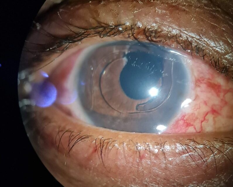
UGH syndrome, standing for uveitis-glaucoma-hyphema syndrome, is a delayed complication of intraocular lens implantation in which mechanical irritation from a malpositioned or poorly sited lens produces the triad the name describes.
It is a classic teaching example of how a mechanical problem, chronic contact between an implant and adjacent uveal tissue, can produce a combination of inflammatory, hemorrhagic, and pressure findings that might otherwise be investigated as three separate, unrelated problems.
Recognizing the triad as a single mechanical process, rather than chasing uveitis, glaucoma, and hyphema independently, is what actually leads to the correct diagnosis and treatment.
UGH syndrome has become less common as lens design and surgical technique have improved, but it has not disappeared entirely, and the underlying principle applies just as directly to any modern lens that ends up malpositioned.
Mechanism
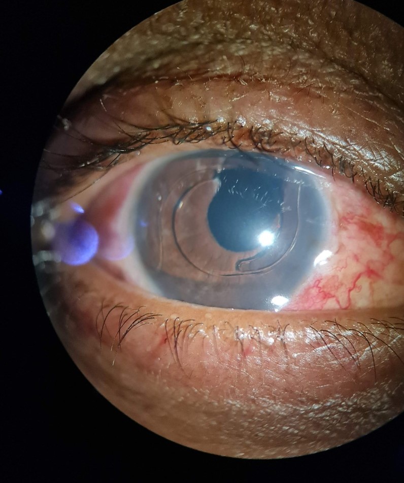
Chronic mechanical chafing occurs when an intraocular lens, most classically an anterior chamber lens or a posterior chamber lens with a haptic malpositioned in the sulcus, repeatedly contacts the iris, ciliary body, or angle structures with eye movement and pupillary changes.
This repetitive contact causes low-grade inflammation, presenting as anterior chamber cell and flare, along with intermittent microhemorrhage from irritated iris or ciliary body vessels, seen clinically as recurrent or persistent hyphema or microhyphema.
Elevated intraocular pressure follows from a combination of inflammatory debris, red blood cells, and pigment obstructing trabecular outflow, compounding the direct mechanical component in some cases where the lens is also physically crowding the angle.
Risk Factors
- Anterior chamber intraocular lenses, historically more associated with UGH syndrome than posterior chamber lenses, given their direct proximity to angle structures
- Sulcus-placed posterior chamber lenses not specifically designed for sulcus fixation, where a haptic can erode against the posterior iris surface over time
- Lens malposition or decentration from any cause, increasing focal contact with adjacent uveal tissue at a specific point
- A history of complicated cataract surgery, including capsular rupture, which increases the likelihood of lens malposition in the first place
A single-piece acrylic lens placed inadvertently in the sulcus, rather than the specific three-piece designs meant for sulcus fixation, is a particularly well-recognized modern-era cause, since the stiffer, flatter haptics of single-piece designs are more prone to chronic posterior iris chafing.
All-fit Slit-Lamp Adapter
Record and share exactly what you see at the slit lamp. One adapter fits any slit lamp or surgical microscope — and any smartphone.
From Choroida — the team behind this siteClinical Presentation
Symptoms and findings typically develop months to years after the original cataract or lens implantation surgery, distinguishing UGH syndrome from the more immediate postoperative inflammation and pressure elevation that can occur in the early recovery period.
Patients may report intermittent blurred vision, floaters from recurrent microhemorrhage, or eye pain, sometimes accompanied by visibly recurrent hyphema noticeable even without a slit lamp.
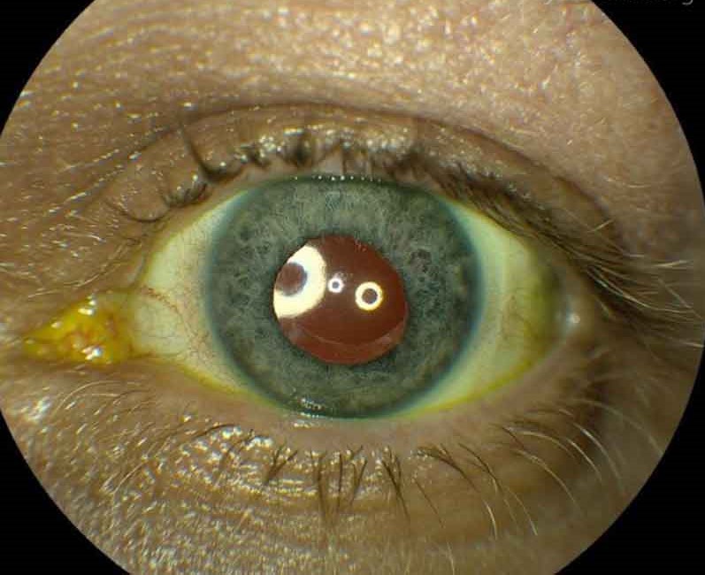
Exam findings include anterior chamber cell and flare, transillumination defects in the iris corresponding to points of chronic lens contact, and elevated intraocular pressure that can range from mild to severe depending on how much outflow obstruction has accumulated.
Diagnostic Evaluation
Careful slit-lamp examination with retroillumination is often the most useful single step, since it can reveal iris transillumination defects that directly localize the point of chronic mechanical contact with the lens.
Gonioscopy assesses the lens position relative to the angle and can reveal a malpositioned haptic directly, while ultrasound biomicroscopy offers a more detailed view of the ciliary sulcus and lens-iris relationship when the diagnosis remains uncertain on exam alone.
Careful review of the original operative report, when available, can clarify what type of lens was implanted and whether any intraoperative complication might explain a subsequently malpositioned implant.
Management
Medical management with topical corticosteroids for inflammation and aqueous suppressants for pressure control can control symptoms temporarily, but it does not address the underlying mechanical cause and recurrence is common once therapy is tapered.
Definitive treatment usually requires surgical repositioning, exchange, or removal of the offending lens, addressing the mechanical irritation at its actual source rather than continuing to treat its downstream inflammatory and hemorrhagic consequences indefinitely.
Careful preoperative planning for the lens exchange, including anticipating capsular support and choosing an appropriate replacement lens type and fixation method, matters considerably for preventing recurrence of the same problem with a new implant.
Because the diagnosis is easy to overlook years after the original surgery, when the patient may no longer connect their new symptoms to an old cataract operation, taking a lens implant history seriously in any unexplained recurrent hyphema or glaucoma is well worth the effort.


Document what you see
Two smartphone imaging tools built for everyday clinic use — one for the slit lamp, one for the fundus.
From Choroida — the team behind this siteReferences
- Ellingson FT. The uveitis-glaucoma-hyphema syndrome associated with the Mark VIII anterior chamber lens implant. Journal of the American Intra-Ocular Implant Society.
- American Academy of Ophthalmology. Basic and Clinical Science Course, Section 11: Lens and Cataract.
- Piette S, Canlas OA, Tran HV, et al. Ultrasound biomicroscopy in uveitis-glaucoma-hyphema syndrome. American Journal of Ophthalmology.
Test yourself
A few questions straight from this article.
-
Which triad of findings gives uveitis-glaucoma-hyphema syndrome its name?
UGH syndrome is a delayed complication of intraocular lens implantation producing uveitis, glaucoma and hyphema together from one mechanical cause. -
What underlying process produces all three findings of UGH syndrome?
The syndrome arises from one mechanical problem: an intraocular lens repeatedly contacting iris, ciliary body or angle structures with eye movement and pupillary change. -
In UGH syndrome, where does the blood of the recurrent hyphema come from?
Repetitive contact causes intermittent microhaemorrhage from irritated iris or ciliary body vessels, seen clinically as recurrent hyphema or microhyphema. -
What raises intraocular pressure in uveitis-glaucoma-hyphema syndrome?
Trabecular outflow is obstructed by inflammatory debris, red blood cells and pigment, sometimes compounded by the lens physically crowding the angle. -
Which intraocular lens placement is a well-recognised modern cause of UGH syndrome?
Single-piece acrylic lenses are not designed for sulcus fixation, and their stiffer, flatter haptics are prone to chronic chafing against the posterior iris. -
How long after lens implantation does UGH syndrome typically declare itself?
The delayed onset, months to years later, separates UGH syndrome from the immediate postoperative inflammation and pressure rise of the early recovery period. -
Which slit-lamp finding localises the point of chronic lens contact in UGH syndrome?
Retroillumination reveals iris transillumination defects that correspond directly to where the implant is chafing against the iris. -
Which imaging gives a detailed view of the lens-iris relationship when UGH syndrome is uncertain?
Ultrasound biomicroscopy shows the ciliary sulcus and the lens-iris relationship in detail when slit-lamp examination and gonioscopy leave the diagnosis unresolved. -
Why does medical therapy alone usually fail to cure UGH syndrome?
Topical steroid and aqueous suppressants control inflammation and pressure only temporarily, because the malpositioned lens keeps chafing once therapy is withdrawn. -
What is the definitive treatment for established uveitis-glaucoma-hyphema syndrome?
Definitive treatment addresses the irritation at its source by repositioning, exchanging or removing the offending implant, with planning for capsular support and fixation to prevent recurrence.
