
Premacular subhyaloid hemorrhage is blood that collects between the posterior hyaloid face and the retina at the macula, and it can drop central vision to hand movements within seconds.
It is often dramatic in appearance, with a dense boat-shaped collection and a flat upper border, and it frequently occurs in otherwise healthy young people after straining.
A single pulse of Nd:YAG laser can release the blood into the vitreous cavity and restore vision within days, but the technique is useful only when the anatomy is right.
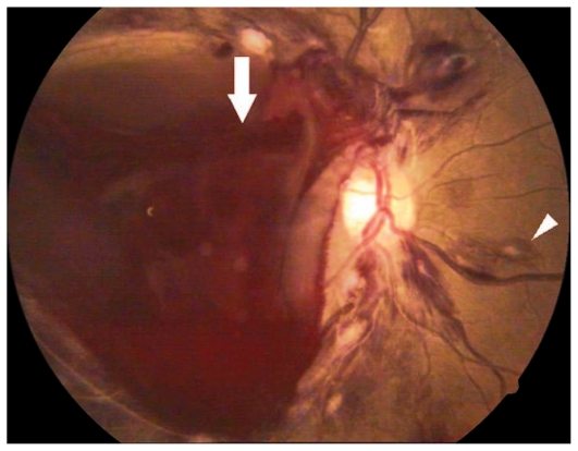
Anatomy of Premacular Subhyaloid Hemorrhage
The hemorrhage lies in one of two potential spaces.
- Subhyaloid (preretinal): between the internal limiting membrane and the posterior hyaloid
- Sub-ILM: between the retina and the internal limiting membrane
A hemorrhage in the subhyaloid space settles by gravity and forms a fluid level with a flat upper margin, and its border shifts with head position.
A sub-ILM hemorrhage has a smoother dome-shaped edge, usually stays in place, and does not form a horizontal level.
Blood originates from superficial retinal capillaries or new vessels that rupture under a sudden rise in venous pressure, or from a disease of the vessels themselves.
The distinction matters because Nd:YAG hyaloidotomy works for a subhyaloid collection and not for one beneath a firmly attached ILM.
Causes
- Valsalva retinopathy after straining, coughing, vomiting, or heavy lifting (see Valsalva retinopathy)
- Proliferative diabetic retinopathy and other neovascular disease
- Retinal vein occlusion (see central retinal vein occlusion)
- Retinal arterial macroaneurysm (see retinal macroaneurysm)
- Terson syndrome after intracranial hemorrhage (see Terson syndrome)
- Blood disorders such as leukemia, thrombocytopenia, and anticoagulation
- Trauma, including abusive head trauma in infants
- Posterior vitreous detachment with retinal vessel avulsion
- Retinal tears or vascular tufts
Valsalva retinopathy in young adults with no other cause is the classic setting for a sudden premacular hemorrhage.
Fundus Explorer Pro
Photograph the retinal findings described here with the phone already in your pocket — 22 D optics and built-in illumination in one handheld unit.
From Choroida — the team behind this siteClinical Features
Patients report a sudden dark or red spot in the central field, sometimes after a specific strain.
Acuity is markedly reduced, and peripheral vision is spared.
Examination shows a round or oval hemorrhage, often about one to three disc diameters, with a fluid level in the subhyaloid form.
The rest of the fundus should be examined carefully, since the cause may be visible elsewhere.
Both eyes should be evaluated, because bilateral hemorrhage points to a systemic cause.
Blood pressure and a bleeding profile should be checked.
OCT shows the cavity beneath the hyaloid or ILM, and it helps decide whether the blood is sub-ILM.
Management Options
Observation
Many hemorrhages clear spontaneously over weeks to months as the blood breaks down and reabsorbs.
Observation is reasonable for small hemorrhages, or when the patient can tolerate the visual loss and the risk of intervention outweighs the benefit.
The disadvantages are prolonged visual loss and the risk of internal limiting membrane changes, epiretinal membrane, and, in children, amblyopia.
Nd:YAG Laser Hyaloidotomy
Nd:YAG laser can perforate the posterior hyaloid and release the blood into the vitreous cavity, where it disperses and clears faster.
The technique:
- Use a contact lens suitable for posterior segment laser
- Start with low pulse energy, and increase gradually until the membrane opens
- Aim at the inferior border of the hemorrhage, well away from the fovea and large vessels, so the blood drains downward and away from the macula
- Confirm a clear defect and a drop in the height of the hemorrhage
In published series, most patients regained good vision within days to weeks after successful drainage.
Reported complications include vitreous hemorrhage, macular hole, retinal detachment, epiretinal membrane, and unrecognized sub-ILM disease that does not drain.
The procedure is best when the hemorrhage is dense, causing visual loss, and clearly subhyaloid, and the patient understands the risks.
Vitrectomy
Vitrectomy is used when laser fails, when the hemorrhage is sub-ILM, or when another indication is present, such as tractional detachment (see pars plana vitrectomy).
ILM peeling may be needed to release blood trapped beneath the membrane.
Treating the Cause
Address the underlying disease.
Diabetic neovascularization needs PRP or anti-VEGF, and macroaneurysms may need laser or injections.
Valsalva retinopathy needs no other treatment beyond avoiding strain.
Differential Diagnosis
A dense red collection over the macula is not always a subhyaloid hemorrhage.
- Sub-ILM hemorrhage, which does not form a fluid level and needs a different approach
- Subretinal or sub-RPE hemorrhage from macroaneurysm or choroidal neovascularization, which is darker and lies beneath the retinal vessels
- Vitreous hemorrhage that has settled inferiorly
- Choroidal melanoma with overlying hemorrhage, when a dark lesion persists after the blood clears
- Retinal vein occlusion with dense macular hemorrhage
OCT and B-scan ultrasonography help separate these, and a repeat examination after clearance identifies any underlying lesion.
Pitfalls
- Treating a sub-ILM collection with Nd:YAG and expecting drainage
- Firing too close to the fovea or over a large retinal vessel
- Overlooking the cause, such as a macroaneurysm or proliferative retinopathy, once the blood has cleared
- Missing the other eye, where a smaller hemorrhage may be present
- Not warning the patient that floaters will increase for a few days after a successful hyaloidotomy
A drained hemorrhage still needs follow-up, since the released blood can settle and later organize.
Prognosis
Visual outcome is generally good in Valsalva retinopathy and in young patients, whether the hemorrhage is drained or observed.
Outcome in diabetic and vascular disease depends on the underlying retinal condition.
Patients should be told to report recurrent bleeding, floaters, or a curtain.


Document what you see
Two smartphone imaging tools built for everyday clinic use — one for the slit lamp, one for the fundus.
From Choroida — the team behind this siteReferences
- Ulbig MW, Mangouritsas G, Rothbacher HH, Hamilton AM, McHugh JD. Long-term results after drainage of premacular subhyaloid hemorrhage into the vitreous with a pulsed Nd:YAG laser. Arch Ophthalmol. 1998;116:1465-1469.
- Durukan AH, Kerimoglu H, Erdurman C, Demirel A, Karagul S. Long-term results of Nd:YAG laser treatment for premacular subhyaloid haemorrhage owing to Valsalva retinopathy. Eye (Lond). 2008;22:214-218.
- Rennie CA, Newman DK, Snead MP, Flanagan DW. Nd:YAG laser treatment for premacular subhyaloid haemorrhage in diabetic retinopathy. Eye (Lond). 2001;15:519-524.
- Mennel S. Subhyaloidal and macular haemorrhage: localisation and treatment strategies. Br J Ophthalmol. 2007;91:850-852.
- Duane TD. Valsalva hemorrhagic retinopathy. Trans Am Ophthalmol Soc. 1972;70:298-313.
