
CASE REPORT
Case of Branch Retinal Artery Occlusion a 16-year-old boy presented having noticed a brief episode of flashing lights followed by an acute inferior hemifield visual loss in the left eye while walking to class. The visual loss persisted, and the event was completely painless.


Past medical history consisted only of migraines which the patient was not experiencing at the time of visual field loss. He denied any history of smoking, illicit drug use, alcohol consumption, or sexually transmitted infections.
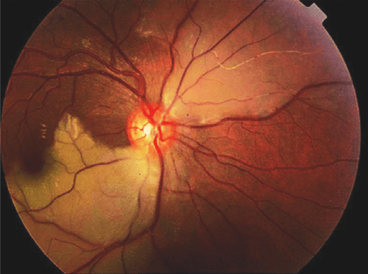
Examination revealed a normal visual acuity of 6/6 in both eyes. A clear inferior altitudinal defect was evident when visual fields were tested to confrontation and a supra-temporal wedge of retinal pallor with associated arterial attenuation was visualized on slit-lamp ophthalmoscope.
Colour retinal photography confirmed evidence of a superior branch retinal artery occlusion (BRAO).
DISEASE of Branch Retinal Artery Occlusion
BRAO, is a common disorder of the ocular vasculature, stems from the occlusion of a branch of the central retinal artery.
BRAO represents 38% of all acute retinal artery obstructions. The resultant hypoperfusion of retinal tissue may result in vision loss.
While frequently described under one heading, two distinct subtypes comprise the condition: permanent BRAO and transient BRAO. More permanent occlusion typically results in more severe visual losses. Transient BRAO bears a better visual prognosis.
MANAGEMENT of Branch Retinal Artery Occlusion
The most critical aspect of managing patients with BRAO is to assess their risk of stroke. Accordingly, all patients should undergo examination by an internist with carotid ultrasound and/or echocardiography as needed.
Referral to a stroke center is appropriate. BRAO often resolves spontaneously, especially those which are of a transient nature. The literature suggests that the presenting VA with BRAO provides a serviceable indication of visual prognosis.
Because prolonged ischemia often produces irreversible damage, and many occurrences of BRAO improve spontaneously, aggressive management in BRAO is not pursued frequently.
would you have the interest to take retinal photography by smart phone?
Fundus photography is superior to fundus analysis as it enables intraocular pathologies to be photo captured and encrypted information to be shared with colleagues and patients.
Recent technologies allow smartphone-based attachments and integrated lens adaptors to transform the smartphone into a portable fundus camera.

More about other disease such as Cherry red spot, Cogan’s syndrome.
REFERENCES
- Mason JO, 3rd, Shah AA, Vail RS, Nixon PA, Ready EL, Kimble JA. Branch retinal artery occlusion: visual prognosis. American journal of ophthalmology. Sep 2008;146(3):455-457.
- Hayreh SS. Ocular vascular occlusive disorders: natural history of visual outcome. Prog Retin Eye Res. Jul 2014;41:1-25.
- Ros MA, Magargal LE, Uram M. Branch retinal-artery obstruction: a review of 201 eyes. Annals of ophthalmology. Mar 1989;21(3):103-107.
- Greco A, De Virgilio A, Gallo A, et al. Susac’s syndrome–pathogenesis, clinical variants and treatment approaches. Autoimmunity reviews. Aug 2014;13(8):814-821
- Tappeiner C, Garweg JG. Retinal vascular occlusion after vitrectomy with retrobulbar anesthesia-observational case series and survey of literature. Graefe’s archive for clinical and experimental ophthalmology = Albrecht von Graefes Archiv fur klinische und experimentelle Ophthalmologie. Dec 2011;249(12):1831-1835.
Read: Retinal imaging by your smartphone and Best 5 ways for Retinal photography.
