

CASE REPORT
Case report of Valsalva Retinopathy a 23 years old pregnant Asian female with 31 weeks of gestation presented with a history of a sudden decrease in vision in her left eye from seven hours prior to the initial visit.

Her clinical problems were initiated after severe retching and vomiting due to the ingestion of ferrous sulfate tablets.
Her previous medical history was unremarkable. Ophthalmic examination showed the best-corrected visual acuity of 20/20 in the right eye and count finger in 2 meters in the left eye. Pupillary response, eye movement, and anterior segment evaluations were normal.
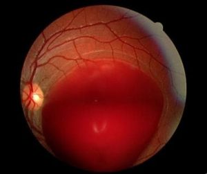
Fundus examination revealed large (10–15 disc diameters) pre-retinal hemorrhage located in the macula.
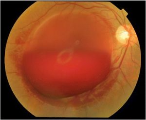
Blood pressure (BP), complete blood count (CBC), coagulation profiles, and fasting blood glucose (FBS) were within the normal ranges. Based on the clinical findings and laboratory studies, the diagnosis of Valsalva retinopathy was confirmed.
DISEASE of Valsalva Retinopathy
Valsalva retinopathy is a preretinal hemorrhage caused by a sudden increase in intrathoracic or intra abdominal pressure. It was first described by Duane in 1972. It usually occurs in an otherwise healthy eye and spontaneously resolves.
It usually occurs in otherwise healthy eyes but may be associated with retinal vascular abnormalities either acquired (diabetic or hypertensive retinopathy) or congenital (retinal telangiectasias and congenital retinal artery tortuosity)
MANAGEMENT of Valsalva Retinopathy
General treatment
Conservative management is the observation of spontaneous resolution, which occurs within weeks to months. Advise patients to avoid anticoagulant drugs and strenuous physical activity.
Surgery
Neodymium: YAG laser or krypton laser membranotomy can be options for large hemorrhage especially if it occurs in the patients only normally functioning eye.
Laser membranotomy disrupts the ILM or posterior hyaloid leading to drainage of the blood into the inferior vitreous cavity, producing a faster resolution.
Complications associated with laser membranotomy include a macular hole, retinal detachment, and epiretinal membrane formation.
In rare cases of premacular hemorrhage, which do not resolve after a reasonable duration of observation, pars plana vitrectomy with removal of ILM and the sub-ILM bleed is an option.
would you have the interest to take retina images by smartphone?
Fundus photography is superior to fundus analysis as it enables intraocular pathologies to be photo captured and encrypted information to be shared with colleagues and patients.
Recent technologies allow smartphone-based attachments and integrated lens adaptors to transform the smartphone into a portable fundus camera.

REFERENCES
- Duane TD. Valsalva hemorrhagic retinopathy. Trans Am Ophthalmol Soc 1972;70:298–313.
- ↑ Kassoff A, Catalano RA, Mehu M. Vitreous hemorrhage and the Valsalva maneuver in proliferative diabetic retinopathy. Retina.1988;8(3):174-6.
- ↑ de Crecchio G, Pacente L, Alfieri MC, Greco GM. Valsalva retinopathy associated with a congenital retinal macrovessel. Arch Ophthalmol. 2000 Jan;118(1):146-7.
- ↑ Tildsley J, Srinivasan S. Valsalva retinopathy. Postgrad Med J 2009;85:110.
- ↑ Agarwall A. Gass’ Atlas of Macular Disease. 5th ed. Elsevier; 2012. Chapter 8, 730-731.
Know about more diseases: Roth Spots, Diabetic retinopathy, Coats disease, Commotio Retinae, Macroaneurysms
