
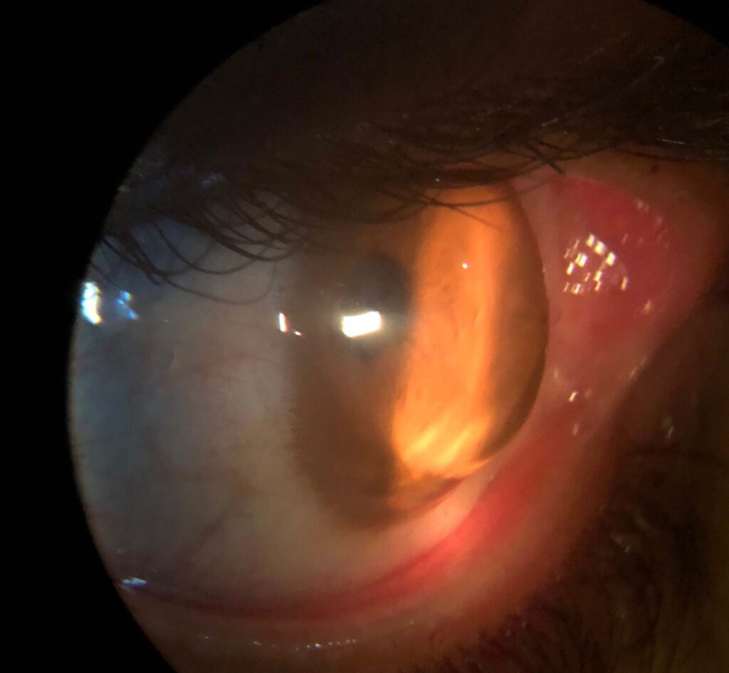
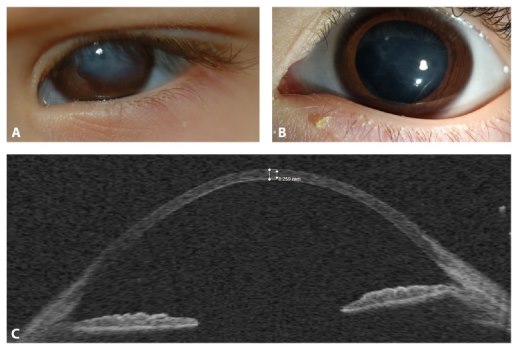
Keratoglobus is a rare, non-inflammatory corneal ectasia in which the entire cornea thins diffusely and balloons into a globular shape, extending out to the limbus rather than staying confined to a central or paracentral cone as in keratoconus. The thinning is most pronounced peripherally, which is the opposite pattern from keratoconus and is what gives the cornea its rounded, oversized appearance.
Most cases are congenital and associated with connective tissue disorders, particularly brittle cornea syndrome and, less consistently, Ehlers-Danlos syndrome. An acquired form exists too, linked to chronic eye rubbing and, historically, to vernal keratoconjunctivitis.
Pathogenesis
Histologically, the corneal stroma in keratoglobus shows thinning to roughly a third of normal thickness, with a reduced number of collagen lamellae and disorganized fibril architecture. Descemet’s membrane is often normal or even thickened, which helps distinguish it from acute hydrops in keratoconus.
In brittle cornea syndrome, mutations in ZNF469 or PRDM5 disrupt collagen and extracellular matrix regulation throughout the body, so the corneal finding is one manifestation of a systemic connective tissue defect rather than an isolated eye disease.
Associated Systemic Findings
- Blue sclerae, from scleral thinning that lets the underlying uvea show through
- Joint hypermobility and skin hyperextensibility
- Hearing loss (in brittle cornea syndrome)
- Hip dysplasia and other skeletal fragility features
- Mitral valve prolapse in some connective tissue overlap syndromes
Recognizing keratoglobus should prompt a search for these features, since the ocular finding is frequently the presenting sign that leads to the systemic diagnosis, especially in a child.
Because the systemic findings can be subtle in isolation, deliberately asking about each of them — hearing, joint mobility, skin texture, prior fractures or dislocations — rather than waiting for them to be volunteered is often what actually surfaces the underlying connective tissue diagnosis.
All-fit Slit-Lamp Adapter
Record and share exactly what you see at the slit lamp. One adapter fits any slit lamp or surgical microscope — and any smartphone.
From Choroida — the team behind this siteClinical Presentation
Vision loss develops gradually from the high, irregular astigmatism that comes with a globally distorted corneal shape. Because the whole cornea is thin, these eyes are also unusually fragile — minor blunt trauma that would be trivial in a normal eye can cause spontaneous corneal or scleral rupture.
Some patients present acutely with hydrops, a sudden break in Descemet’s membrane that lets aqueous flood into the stroma and causes acute pain, photophobia, and marked corneal edema. This is less common than in keratoconus but tends to be more severe when it happens, given the extent of pre-existing thinning.
Differential Diagnosis
- Keratoconus — central/paracentral cone, relatively preserved peripheral cornea
- Pellucid marginal degeneration — inferior peripheral thinning band, “kissing dove” topography
- Megalocornea — enlarged but structurally normal-thickness cornea
- Congenital glaucoma (buphthalmos) — enlarged globe from elevated IOP, Haab’s striae
- Terrien’s marginal degeneration — peripheral thinning, usually superior, in older adults
Diagnostic Evaluation
Slit-lamp examination shows the diffusely thin, domed cornea extending to the limbus without the sharp demarcation typical of keratoconus. Corneal topography and tomography (Scheimpflug imaging) quantify the pattern of thinning and steepening across the whole cornea rather than a focal apex, and pachymetry documents just how thin the tissue is — often under 400 microns, sometimes far less.
Genetic testing is useful when brittle cornea syndrome is suspected, both to confirm the diagnosis and to guide counseling for the family, since it is autosomal recessive. Axial length and corneal diameter measurements help distinguish keratoglobus from buphthalmos, where intraocular pressure — not just corneal shape — is the primary abnormality.
Management
Rigid gas-permeable or scleral contact lenses can improve vision by masking irregular astigmatism, but fitting is difficult given the globular shape and fragility of the cornea, and lens wear itself carries a real risk of mechanical trauma to tissue this thin. Protective eyewear is worth discussing early, since these patients are at genuine risk of globe rupture from impacts that most people would shrug off, and this discussion should extend to sports and occupational activity choices, not just everyday incidental risk.
Corneal transplantation is technically demanding in keratoglobus because there is so little healthy peripheral tissue to support graft-host apposition; large-diameter penetrating keratoplasty or epikeratoplasty has been used, but outcomes are less predictable than in keratoconus and rejection or wound dehiscence risk is higher given the abnormal collagen. Some surgeons favor staged approaches — tectonic patch grafts to reinforce weak areas before attempting an optical procedure — given how technically demanding a single-stage definitive repair can be in this fragile tissue.
Complications and Prognosis
Spontaneous or trauma-related perforation is the complication that most changes management, since these patients need to understand that even modest impact — a fall, a poorly aimed ball, rubbing the eye vigorously — can rupture the globe. Acute hydrops, when it occurs, usually resolves over weeks to months as the endothelium re-establishes its pump function, though it can leave residual scarring that further affects visual quality.
Visual prognosis depends heavily on the severity of the ectasia and whether the systemic disorder is otherwise well managed. Genetic counseling matters for families with brittle cornea syndrome given its recessive inheritance and multisystem impact, and coordinated care across ophthalmology, genetics, and, depending on the specific systemic findings present, orthopedics or audiology reflects the genuinely multisystem nature of this condition.


Document what you see
Two smartphone imaging tools built for everyday clinic use — one for the slit lamp, one for the fundus.
From Choroida — the team behind this siteReferences
- Al-Hussain H, et al. Brittle cornea syndrome and its delineation from the kyphoscoliotic type of Ehlers-Danlos syndrome. American Journal of Medical Genetics.
- Abu A, et al. Mutation in PRDM5 in brittle cornea syndrome. American Journal of Human Genetics.
- Meire FM. Keratoglobus. In: Krachmer JH, Mannis MJ, Holland EJ, eds. Cornea.
- Cameron JA. Keratoglobus. Cornea.
- Rathi VM, Vyas SP, Sangwan VS. Keratoglobus. Indian Journal of Ophthalmology.
Test yourself
A few questions straight from this article.
-
In keratoglobus, where is the corneal thinning most pronounced?
This peripheral-maximum pattern is the opposite of keratoconus and is what gives the cornea its rounded, globular, oversized appearance. -
What does corneal histology show in keratoglobus?
Collagen lamellae are reduced in number with disorganized fibril architecture, while Descemet's membrane is often normal or even thickened. -
Mutations in which genes underlie brittle cornea syndrome with keratoglobus?
These disrupt collagen and extracellular matrix regulation throughout the body, so the cornea is one manifestation of a systemic, autosomal recessive connective tissue defect. -
What causes the blue sclerae associated with keratoglobus in connective tissue disease?
Blue sclerae sit alongside joint hypermobility, skin hyperextensibility, hearing loss and hip dysplasia as clues to the underlying systemic disorder. -
What corneal thickness does pachymetry typically document in keratoglobus?
Pachymetry records how thin the tissue truly is, often under 400 microns and sometimes far less, across the whole cornea rather than at a focal apex. -
Which feature distinguishes buphthalmos from keratoglobus?
In buphthalmos the intraocular pressure, not corneal shape alone, is the primary abnormality; axial length and corneal diameter help separate the two. -
Which corneal ectasia shows an inferior peripheral thinning band with a kissing dove topography?
Keratoglobus thins diffusely to the limbus, whereas pellucid marginal degeneration confines its thinning to an inferior peripheral band. -
What should be discussed early with every patient who has keratoglobus?
These eyes can rupture from impacts most people would shrug off, so the conversation must cover sport and occupation, not just incidental everyday risk. -
Why is corneal transplantation technically demanding in keratoglobus?
Large-diameter penetrating keratoplasty or epikeratoplasty has been used, but rejection and wound dehiscence risk is higher because the collagen is abnormal. -
What is the usual course of acute hydrops occurring in keratoglobus?
Hydrops resolves as the endothelium re-establishes its pump function, though residual scarring can further degrade visual quality.

Writes and reviews the clinical articles on Educate. Every post is checked against peer-reviewed sources before it goes live.


