
CASE REPORT
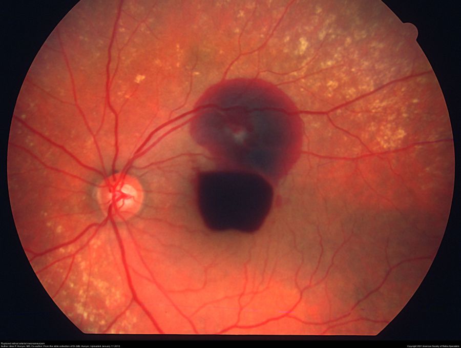
A 62-year-old female experienced sudden visual loss in her left eye for approximately two weeks. The BCVA was limited to counting fingers in the left eye. Dilated fundus examination showed preretinal hemorrhage in the macula area.
OCT scanning of the fovea showed markedly increased retinal thickness. Considering the dense hemorrhage, we only conducted an ICGA examination.
The results showed hyper fluorescence at the inferior temporal area of the edge of the dark area. A diagnosis of retinal arterial macroaneurysm was made.
Disease
Retinal arterial macroaneurysms are acquired, focal dilations of retinal arterial branches (mostly second-order retinal arterioles) that can be classified as hemorrhagic or exudative.
Macroaneurysms range from 100 to 250μm in diameter and are most often found in the temporal retina, along the supero-temporal arteriole. Associated findings include capillary telangiectasias, vascular remodeling, and retinal edema.
While,
the visual prognosis is generally good, vision loss can occur from macular edema, end-arteriole occlusion from thrombosis, or hemorrhage due to rupture of the aneurysm.
Diagnosis is made on clinical exam and via imaging modalities such as fluorescein angiography (FA) and spectral-domain optical coherence tomography (SD-OCT), and treatment is traditionally observation, focal laser photocoagulation, or surgery.
More recently, the use of anti-vascular endothelial growth factor (VEGF) intravitreal injections has been suggested but not extensively studied as a possible treatment modality.
Fundus Explorer Pro
Photograph the retinal findings described here with the phone already in your pocket — 22 D optics and built-in illumination in one handheld unit.
From Choroida — the team behind this siteEtiology and Pathophysiology
Vessel wall thickening and the presence of fibrin and foamy macrophages upon histological inspection in retinal arterial macroaneurysms was described to be similar to that seen in age-related arteriosclerotic changes in the vessel walls throughout the body.
The localized ischemia due to focal embolic damage to the arterial walls from vascular disease can cause increased intimal collagen remodeling and increased permeability of vessels, thus predisposing the vessel to dilation.
Pathology
Linear breaks found in arterial walls on histopathology lead to a round or fusiform dilation of the arteriolar vessel wall. This weakened arteriolar wall can rupture and cause hemorrhage and exudation at any level of the retina.

Epidemiology
Studies have reported on the incidence of arterial macroaneurysms as being 1 case per 9000 primarily.
The majority of those affected are elderly and female, and there is a strong association with hypertension (seen in 75% of patients). In hemorrhagic macroaneurysms, 10% of cases are bilateral.
Primary prevention
Control of hypertension and arteriolar sclerosis risk factors are the mainstay of prevention.
Diagnosis
Fluorescein angiography can help visualize macroaneurysms which typically fill in the early arterial phase and stain the vessel walls. Possible leakage may be found in later phases.
History
Most commonly, retinal arterial macroaneurysm (RAM) is found incidentally on funduscopic examination in an asymptomatic patient.
However, patients may complain of acute vision loss due to macular edema or hemorrhage. Metamorphopsia or decreased vision is most commonly encountered when changes due to macroaneurysms (hemorrhage or edema) affect the fovea.
Also presenting complaints may be floaters due to vitreous hemorrhage. Typically patients will have a history of hypertension.
Physical Examination and Retinal imaging
Dilated fundus examination may enable visualization of an out-pocketing of the arterial wall, but in the setting of hemorrhage and/or exudation, it may be necessary to use imaging modalities such as FA and SD-OCT to confirm the diagnosis.
Round or fusiform dilation of a retinal arteriole is usually seen within a third-degree branch of one of the four main arcade arteries.
The most common location for asymptomatic macroaneurysm is from a branch of the superotemporal arcade.
The aneurysm appears as a pulsating round or ovoid pooling of dye on FA, with filling either in the middle to a late phase in saccular (sac-like) aneurysms or in the early phase in fusiformic (spindle-shaped) aneurysms.
SD-OCT can be helpful in quantifying the exudates in the exudative-type macroaneurysm.
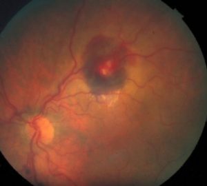
Retinal arterial macroaneurysm with subretinal and preretinal hemorrhage.
Differential Diagnosis
While the presence of a retinal arterial macroaneurysm can be visualized on fundus examination, certain disease processes can present similarly such as Coat’s disease and von Hippel-Lindau disease.
Macroaneurysms can also occur secondary to a branch retinal vein occlusion, diabetic retinopathy, radiation retinopathy, and retinal arteritis, in which case the underlying cause should be teased out and treated.
RAM is one of the few causes of hemorrhage at multiple levels (preretinal, retinal, and sibretinal hemorrhage). Other causes include anemic/leukemic retinopathy and trauma (shaken baby syndrome).
Management
There are currently no approved guidelines for the management of macroaneurysms. Most macroaneurysms resolve spontaneously and can be observed.
In all patients with this diagnosis, a systematic workup for hypertension and systemic vascular disease should be pursued.
Complicated retinal arterial macroaneurysms may be directly treated with moderate-intensity laser photocoagulation with two to three rows of large-spot-size (200-500μm) immediately adjacent to the macroaneurysm, especially if the visual function is threatened due to increasing edema.
This treatment course is controversial, as some studies have demonstrated a significant decrease in visual acuity in post-laser eyes and the possible occurrence of branch retinal arterial occlusion.
Cases of submacular hemorrhage can be treated with a pneumatic displacement of the clot with or without tissue plasminogen activator, as hemorrhage is toxic to the retina and ellipsoid zone.
In cases of premacular hemorrhage, YAG laser can be used to perform a posterior hyaloidotomy and release the hemorrhage into the vitreous for faster clearance.
Prognosis
The majority of eyes with the diagnosis of arterial macroaneurysm have good visual prognoses and spontaneous regression.
In general,
patients with a hemorrhagic macroaneurysm have a better visual prognosis than those with an exudative macroaneurysm. Patients with persistent macular edema or subretinal hemorrhage were found to have the worst prognosis.
Fundus photography is superior to fundus analysis as it enables intraocular pathologies to be photo captured and encrypted information to be shared with colleagues and patients.
Recent technologies allow smartphone-based attachments and integrated lens adaptors to transform the smartphone into a portable fundus camera.
Read about:Valsalva Retinopathy, Best 5 ways for retinal photography, Birdshot Retinochoroidopathy


Document what you see
Two smartphone imaging tools built for everyday clinic use — one for the slit lamp, one for the fundus.
From Choroida — the team behind this siteReferences
- Lee EK, Woo SJ, Ahn J, Park KH. Morphologic characteristics of retinal arterial macroaneurysm and its regression pattern on spectral-domain optical coherence tomography. Retina 2011;31(10):2095-2101.
- Moorsavi RA, Fong KC, CHopdar A. Retinal artery macroaneurysms: clinical and fluorescein angiographic features in 34 patients. Eye (Lond) 2006;20(9):1011-1020.
- Fichte C, Streeten BW, Friedman AH. A histopathologic study of retinal arterial aneurysms. Am J Ophthalmol 1978;85:509-518.
- Xu L, Wang Y, Jonas JB. Frequency of retinal macroaneurysms in adult Chinese: the Beijing Eye Study. Br J Ophthalmol 2007;91:840-841.
- Panton RW, Goldberg MF, Farber MD (1990): Retinal arterial macroaneurysms: risk factors and natural history. Br J Ophthalmol 74: 595-600.
- Rabb MF, Gagliano DA, Teske MP. Retinal arterial macroaneurysms. Surv Ophthalmol 1988;33(2):73-96.
- Lewis RA, Norton EW, Gass JD (1976): Acquired arterial macroaneurysms of the retina. Br J Ophthalmol 60:21-30.
- Theodossaidis PG, Emfietzoglou I, Sfikakis PP, Panagiotidis D, Grigoropoulous VG., Theodossiadis GP (2005): simultaneous bilateral visual loss caused by rupture of retinal arterial macroaneurysm in a hypertensive patient. Acta Ophthalmol cand 83:120-122.
Test yourself
A few questions straight from this article.
-
Retinal arterial macroaneurysms are acquired focal dilations of which vessels?
They are acquired focal dilations of retinal arterial branches, most often second-order retinal arterioles, and are classified as haemorrhagic or exudative. -
What diameter range is typical for a retinal arterial macroaneurysm?
Macroaneurysms measure roughly 100-250 micrometres across, large enough to be seen as a discrete out-pocketing of the arteriolar wall. -
Which systemic disease is present in about 75% of patients with retinal arterial macroaneurysm?
Most affected patients are elderly women, and hypertension is found in roughly three quarters of them, so blood pressure control is the mainstay of prevention. -
Where is an asymptomatic retinal arterial macroaneurysm most commonly found?
Macroaneurysms favour the temporal retina, classically arising from a third-degree branch of the superotemporal arcade artery. -
What do the arterial walls show histologically in retinal arterial macroaneurysm?
The thickened wall containing fibrin and foamy macrophages mirrors age-related arteriosclerotic change elsewhere in the body, and linear wall breaks then allow dilation. -
How does a retinal arterial macroaneurysm typically behave on fluorescein angiography?
Macroaneurysms usually fill in the early arterial phase and stain the vessel wall, with leakage sometimes appearing in later frames. -
Which conditions can mimic retinal arterial macroaneurysm on fundus examination?
Coats disease and von Hippel-Lindau disease can look similar on fundoscopy; macroaneurysms may also arise secondary to vein occlusion or diabetic retinopathy. -
What haemorrhage pattern makes retinal arterial macroaneurysm distinctive?
It is one of the few causes of haemorrhage at multiple retinal levels at once; anaemic or leukaemic retinopathy and trauma are the other classic causes. -
What is the usual initial management of an uncomplicated retinal arterial macroaneurysm?
No approved treatment guidelines exist and most macroaneurysms regress on their own, so observation plus a workup for hypertension and vascular disease is standard. -
How may submacular haemorrhage from a retinal arterial macroaneurysm be treated?
Subretinal blood is toxic to the retina and ellipsoid zone, so the clot may be displaced pneumatically, with or without tissue plasminogen activator.

Writes and reviews the clinical articles on Educate. Every post is checked against peer-reviewed sources before it goes live.


